

Volume 12, Issue 1 (June 2026)
Elderly Health Journal 2026, 12(1): 34-45 |
Back to browse issues page



Download citation:
BibTeX | RIS | EndNote | Medlars | ProCite | Reference Manager | RefWorks
Send citation to:


BibTeX | RIS | EndNote | Medlars | ProCite | Reference Manager | RefWorks
Send citation to:
Janghorbani F, Taghian F, Jalali Dehkordi K. The Effect of an Eight-Week Low-Volume High-Intensity Interval Training Compared to Moderate-Intensity Continuous Training on Liver Enzymes and Anthropometric Indices: A Comparison of Obese and Lean Men with Non-Alcoholic Fatty Liver Disease. Elderly Health Journal 2026; 12 (1) :34-45
URL: http://ehj.ssu.ac.ir/article-1-371-en.html
URL: http://ehj.ssu.ac.ir/article-1-371-en.html


Department of Sports Physiology, Isf.C., Islamic Azad University, Isfahan, Iran , ft.taghian@gmail.com
Keywords: Non-Alcoholic Fatty Liver Disease, Body Mass Index, Waist Hip Ratio, Alanine Transaminase, Aspartate Aminotransferases
Full-Text [PDF 543 kb]
(149 Downloads)
| Abstract (HTML) (496 Views)
Full-Text: (122 Views)
The Effect of an Eight-Week Low-Volume High-Intensity Interval Training Compared to Moderate-Intensity Continuous Training on Liver Enzymes and Anthropometric Indices: A Comparison of Obese and Lean Men with Non-Alcoholic Fatty Liver Disease
Fatemeh Janghorbani 1, Farzaneh Taghian 1*, Khosro Jalali Dehkordi 1
Article history
A B S T R A C T
Keywords: Non-Alcoholic Fatty Liver Disease, Body Mass Index, Waist Hip Ratio, Alanine Transaminase, Aspartate Aminotransferases
Table 1. High intensity interval training (HIIT) protocol over the 8-week intervention
Table 2. Moderate intensity continuous training (MICT) protocol over the 8-week intervention
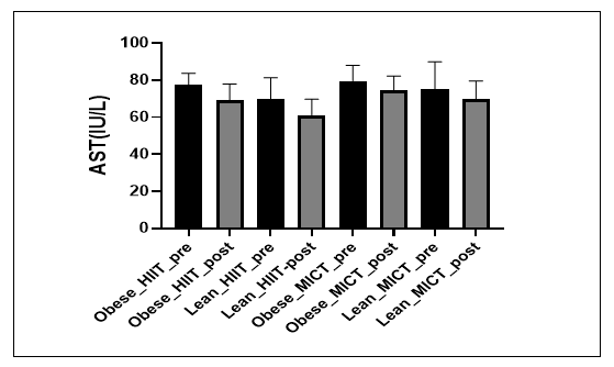
Figure 1. Changes in serum aspartate aminotransferase (AST) levels in obese and normal weight/lean men with NAFLD following 8 weeks of high intensity interval training (HIIT) and moderate intensity continuous training (MICT).
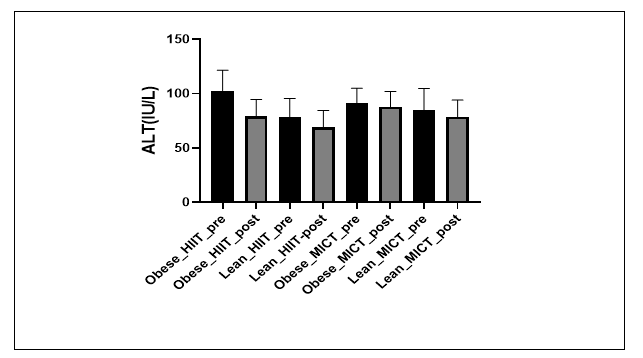
Figure 2. Changes in serum alanine aminotransferase (ALT) levels in obese and normal weight/lean men with NAFLD following 8 weeks of high Figure 2. Changes in serum alanine aminotransferase (ALT) levels in obese and normal weight/lean men with NAFLD following 8 weeks of high intensity interval training (HIIT) and moderate intensity continuous training (MICT).
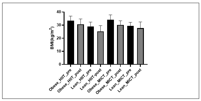
Figure 3. Pre and post intervention changes in body mass index (BMI) in obese and normal weight/lean men undergoing high intensity interval training (HIIT) and moderate intensity continuous training (MICT).
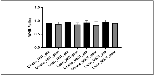
Figure 4. Changes in waist to hip ratio (WHR) following 8 weeks of high intensity interval training (HIIT) and moderate intensity continuous training (MICT) in obese and normal weight/lean men with NAFLD.
Table 3. Mean ± SD of ALT, AST, BMI, and WHR before and after the 8-week exercise intervention in obese and lean men with NAFLD
Data are presented as mean ± standard deviation (SD).
Between group comparisons were conducted using analysis of covariance (ANCOVA), with baseline values entered as covariates.
Partial eta‑squared (partial η²) was used as a measure of effect size, with values of 0.01, 0.06, and 0.14 interpreted as small, medium, and large effects, respectively.
HIIT = high‑intensity interval training; MICT = moderate intensity continuous training;
BMI = body mass index; WHR = waist‑to‑hip ratio;
ALT = alanine aminotransferase; AST = aspartate aminotransferase.
Fatemeh Janghorbani 1, Farzaneh Taghian 1*, Khosro Jalali Dehkordi 1
- Department of Sports Physiology, Isf.C., Islamic Azad University, Isfahan, Iran
Article history
Received 19 Nov 2025
Accepted 17 Dec 2025
A B S T R A C T
Introduction: Nonalcoholic fatty liver disease (NAFLD) is a prevalent metabolic liver disorder associated with insulin resistance, excess adiposity, and increased cardiovascular risk. Although obesity is a major risk factor, many individuals with NAFLD are lean, and their responses to exercise remain unclear. This study compared the effects of eight weeks of high-intensity interval training (HIIT) and moderate-intensity continuous training (MICT) on liver enzymes and anthropometric indices in obese and lean men with NAFLD.
Methods: In this quasi-experimental study, 56 men with ultrasound confirmed NAFLD (grade ≥ 2) were randomly allocated to four groups: obese + HIIT, obese + MICT, lean + HIIT, and lean + MICT. Participants completed supervised exercise sessions three times per week for eight weeks. Serum alanine aminotransferase (ALT), aspartate aminotransferase (AST), body mass index (BMI), and waist to hip ratio (WHR) were measured before and after the intervention. Between group differences were analyzed using analysis of covariance (ANCOVA), with baseline values as covariates. Effect sizes were reported using partial eta squared (η²).
Results: After adjustment for baseline values, no significant between-group differences were observed for ALT or AST, although both enzymes showed numerical reductions in all groups. Significant between-group differences were found for anthropometric outcomes. HIIT produced greater reductions in BMI (p < 0.001, partial η² = 0.38) and WHR (p < 0.001, partial η² = 0.29) than MICT, with the greatest improvements observed in obese participants.
Conclusion: An eight-week HIIT program was more effective than MICT in improving body composition, particularly overall and central adiposity, in men with NAFLD. Although exercise was associated with favorable trends in liver enzyme levels, these changes did not reach statistical significance between groups. These findings support HIIT as a time efficient strategy for improving anthropometric outcomes in men with NAFLD.
Methods: In this quasi-experimental study, 56 men with ultrasound confirmed NAFLD (grade ≥ 2) were randomly allocated to four groups: obese + HIIT, obese + MICT, lean + HIIT, and lean + MICT. Participants completed supervised exercise sessions three times per week for eight weeks. Serum alanine aminotransferase (ALT), aspartate aminotransferase (AST), body mass index (BMI), and waist to hip ratio (WHR) were measured before and after the intervention. Between group differences were analyzed using analysis of covariance (ANCOVA), with baseline values as covariates. Effect sizes were reported using partial eta squared (η²).
Results: After adjustment for baseline values, no significant between-group differences were observed for ALT or AST, although both enzymes showed numerical reductions in all groups. Significant between-group differences were found for anthropometric outcomes. HIIT produced greater reductions in BMI (p < 0.001, partial η² = 0.38) and WHR (p < 0.001, partial η² = 0.29) than MICT, with the greatest improvements observed in obese participants.
Conclusion: An eight-week HIIT program was more effective than MICT in improving body composition, particularly overall and central adiposity, in men with NAFLD. Although exercise was associated with favorable trends in liver enzyme levels, these changes did not reach statistical significance between groups. These findings support HIIT as a time efficient strategy for improving anthropometric outcomes in men with NAFLD.
Keywords: Non-Alcoholic Fatty Liver Disease, Body Mass Index, Waist Hip Ratio, Alanine Transaminase, Aspartate Aminotransferases
Copyright © 2026 The Author(s). This is an open-access article distributed under the terms of the Creative Commons Attribution-Non Commercial 4.0 International License (http://creativecommons.org/licenses/by-nc/4.0/) which permits copy and redistribute the material just in noncommercial usages, provided the original work is properly cite.
Introduction
Non-alcoholic fatty liver disease (NAFLD) has emerged as the most prevalent chronic liver disorder in recent decades, encompassing a broad spectrum ranging from simple steatosis to nonalcoholic steatohepatitis (NASH), fibrosis, and eventually cirrhosis or hepatocellular carcinoma. Recent global reviews and systematic analyses estimate the prevalence of NAFLD to be approximately 30–32%, and its increasing trend, which parallels the global epidemics of obesity and metabolic syndrome, imposes a substantial and growing burden on healthcare systems worldwide and on economic resources. The fact that NAFLD can lead to systemic complications such as cardiovascular diseases and type 2 diabetes highlights the importance of preventive research and non-pharmacological therapeutic interventions (1).
From a pathophysiological perspective, NAFLD is a multifactorial disease in which insulin resistance, lipid metabolism dysregulation, mitochondrial dysfunction, and inflammatory responses play pivotal roles (1). Obesity particularly central or visceral adiposity-is recognized as one of the most critical underlying factors in the development and progression of NAFLD (2). Simple clinical indicators such as body mass index (BMI) and waist-to-hip ratio (WHR) have shown a positive correlation with the degree of hepatic fat accumulation and biochemical markers of liver function in numerous studies (1, 2).
In certain cases, WHR or other indices of fat distribution may serve as stronger predictors of NAFLD risk even among individuals with normal BMI; therefore, the simultaneous assessment of both fat quantity and distribution is essential in interventional research (2). Although obesity is a major contributing factor to NAFLD, approximately 20% of patients with this condition are lean (3). NAFLD in individuals with normal body weight defined as a BMI below 23 kg/m² in Asians and below 25 kg/m² in other ethnic groups is referred to as lean NAFLD. This phenotype is generally considered less severe than NAFLD associated with higher BMI levels. Because lean NAFLD patients typically exhibit more favorable metabolic profiles and histological features, it is commonly assumed that this lean subgroup experiences a relatively benign clinical course compared with their overweight or obese counterparts (4).
A landmark study by Cruz et al., was the first to demonstrate that individuals with lean NAFLD exhibit a higher mortality risk than their overweight or obese counterparts (5). Subsequently, several studies investigating mortality risk associated with lean NAFLD have yielded controversial findings (6-9). Because lean NAFLD patients are generally characterized by fewer metabolic complications, it has long been assumed that they have a lower risk of developing cardiovascular disease (CVD) compared with individuals with higher BMI (10). Nevertheless, a prospective cohort study conducted in the United States demonstrated that lean individuals with NAFLD were at a comparable risk of developing metabolic disorders and CVD as their obese counterparts during long term follow up (10).
Obesity is a major risk factor for metabolic disorders, primarily due to adipocyte dysfunction, as adipose tissue plays a crucial role in lipid storage and endocrine regulation. In obese individuals, adipocytes become hypertrophic and metabolically impaired, leading to altered secretion of adipokines such as leptin and adiponectin. Obesity, characterized by abnormal fat accumulation, is both a risk factor and an aggravating contributor to NAFLD, as well as a well-established risk factor for type II diabetes. Adipose tissue dysfunction promotes chronic low-grade inflammation and ectopic lipid accumulation, thereby contributing to systemic insulin resistance (1, 11).
Insulin resistance in obesity is characterized by impairments in insulin signaling pathways, particularly within adipose tissue and the liver. Physiologically, insulin regulates blood glucose levels, adipose tissue lipolysis, and hepatic synthesis of very low-density lipoproteins (VLDL). In insulin-resistant individuals, increased release of free fatty acids (FFAs) from adipose tissue, along with enhanced hepatic uptake of FFAs, leads to their conversion into hepatic triglycerides, thereby contributing to hepatic steatosis (12). At the cellular level, elevated fatty acid concentrations induce endoplasmic reticulum stress, which inhibits the secretion of apolipoprotein B100 and promotes the progression of steatosis. Furthermore, evidence suggests that hepatic steatosis itself may represent the initial event triggering insulin resistance in lean individuals (13).
Lifestyle modification, particularly weight reduction and structured physical activity, forms the cornerstone of clinical recommendations for managing NAFLD. Evidence from randomized controlled trials and meta analyses indicates that aerobic, resistance, or combined exercise programs can reduce hepatic fat, improve insulin sensitivity parameters, and, in some cases, decrease serum transaminase levels (ALT and AST); however, the optimal type, intensity, and volume of exercise remain under investigation (14, 15). Recent meta-analyses suggest that both moderate-intensity continuous training (MICT) and high intensity interval training (HIIT) can lead to significant reductions in hepatic fat, yet evidence regarding the superiority of one approach over the other is inconsistent, highlighting the need for studies with direct comparative designs and larger sample sizes (14, 16).
The benefits of regular physical activity in reducing the risk of certain extrahepatic lifestyle related cancers are well established. The profound effects of exercise on cardiometabolic risk, vascular health, and cardiorespiratory fitness have been extensively documented across a wide range of clinical populations (17). Given evidence suggesting that participants’ body weight may influence the effectiveness of exercise on clinical markers of NAFLD, patient responses could vary according to body fat levels. Furthermore, the capacity to perform physical activity may be affected by excess body weight, which in turn can impact metabolic responses and adaptive outcomes. While further research is needed to clarify whether the degree of obesity influences the effectiveness of exercise on clinical markers of NAFLD, it has been established that weight reduction is associated with significant pathophysiological improvements, particularly enhanced insulin sensitivity, decreased hepatic free fatty acids, attenuation of inflammatory mechanisms, and improved ALT and AST levels (18). In this context, Katsagoni et al., reported that the effects of exercise on AST and ALT are dependent on weight loss. Moderate-intensity aerobic training of higher volume (over 180 minutes per week) has been shown to be more effective in reducing intrahepatic triglycerides than moderate intensity aerobic exercise of lower volume (120–180 minutes per week) (19).
Regular physical activity has been shown to enhance mitochondrial oxidative capacity and increase mitochondrial content, which is associated with improvements in cardiorespiratory fitness (20). Indeed, cardiorespiratory fitness is inversely correlated with hepatic steatosis, and improvements in fitness have been independently associated with reductions in hepatic fat (21). Given that cardiorespiratory fitness has been demonstrated to improve to a similar extent following both high-intensity interval training (HIIT) and traditional moderate intensity continuous training, this may partly explain why HIIT, despite lower energy expenditure, results in comparable improvements in hepatic steatosis relative to moderate-intensity continuous exercise (22).
The comparison between HIIT and MICT is noteworthy from both clinical and practical perspectives. Firstly, HIIT is considered an attractive option for time-constrained patients due to its shorter time commitment, capacity to induce post exercise metabolic adaptations (excess post exercise oxygen consumption, EPOC), and enhancement of fat oxidation. Secondly, MICT may offer greater practical accessibility owing to its ease of implementation and broader acceptability among participants (20, 22).
Recent reviews and meta analyses indicate that, although both approaches are effective in reducing hepatic fat, statistical differences in enzymatic outcomes or histological liver changes between the two methods depend on intervention duration, controlled intensity, and study specific procedural mechanisms. For instance, randomized and re-examined trials, such as those by Sabbagh et al., have demonstrated that low volume aerobic interventions can significantly reduce hepatic fat, and pooled comparative studies have further explored the relative effects of HIIT and MICT (16, 23).
Despite the growing body of evidence, several methodological limitations in the current literature restrict the generalizability of findings. Firstly, many studies have focused primarily on obese populations, and systematic comparative investigations of exercise effects across different body composition groups (e.g., obese versus normal weight/lean) remain scarce. This gap hinders the determination of whether enzymatic responses and changes in BMI or WHR are intrinsically influenced by baseline body composition. Secondly, variations in exercise protocols (duration and intensity), the presence or absence of precise dietary control, and the timing of blood sampling (e.g., 24 hour versus 72 hour post exercise) can introduce inconsistencies across studies and complicate data interpretation (2, 24). Recent meta-analyses have recommended that future studies adopt standardized protocols, appropriate blood sampling schedules to eliminate acute exercise effects, and comparative clinical designs to enable robust and symmetrical analyses (2, 16, 24).
From a mechanistic perspective, the exercise-induced reduction of hepatic fat involves improvements in insulin sensitivity, enhanced skeletal muscle and hepatic fat oxidation, attenuation of systemic inflammation, and modulation of gene expression and metabolic pathways associated with lipid accumulation. Evidence indicates that regular exercise can decrease hepatic lipid burden by reducing dysregulated lipolysis in visceral adipose tissue and improving hepatic metabolic function. These mechanisms have been supported in both animal models and human studies; however, exercise intensity and modality may influence the magnitude and rate of these adaptations, highlighting the need for more detailed investigations of metabolic markers and biomolecular pathways (14, 15).
Given the aforementioned gaps and uncertainties, studies specifically designed to examine differential responses to HIIT and MICT interventions in individuals with varying body compositions (obese versus normal weight/lean) are of critical importance. Such studies should employ standardized exercise protocols, minimal dietary control (e.g., dietary recall), and appropriate blood sampling schedules to mitigate transient and carryover exercise effects, thereby enabling meaningful and interpretable reporting of enzymatic (ALT, AST) and anthropometric (BMI, WHR) outcomes (2, 16, 24). Accordingly, the present study, conducted as an 8-week, four-group intervention (obese + HIIT, obese + MICT, lean + HIIT, lean + MICT), represents an effort to address these gaps and provide practical evidence for tailoring and optimizing exercise programs in patients with NAFLD.
The primary aim of this study is to evaluate and compare the effects of an 8 week HIIT versus MICT program on hepatic enzymes (ALT and AST) and anthropometric indices (BMI and WHR) in men with NAFLD grade ≥ 2, with a particular focus on stratified analyses according to body composition (obese versus normal weight/lean). It is anticipated that the findings of this investigation will provide critical evidence to inform the selection of exercise type and intensity tailored to patients’ body composition, thereby offering clearer clinical guidance for non-pharmacological interventions in NAFLD.
Methods
Study design
This study was conducted as quasi-experimental research with a pretest–posttest design involving four parallel groups. The intervention period lasted eight weeks. Participants were allocated to one of the following groups: obese + high-intensity interval training (HIIT); obese + moderate-intensity continuous training (MICT); normal-weight/lean + HIIT; or normal-weight/lean + MICT.
Participants and sample size
The study population consisted of men diagnosed with NAFLD grade 2 or 3 who were referred to outpatient clinics in Isfahan County and Isfahan Health Town, Iran. Participants were recruited consecutively over [specific period], and informed consent was obtained prior to enrollment. Sample size estimation was performed using G*Power software (version 3.1.9.2), with a statistical power of 0.80, a significance level of 0.05, and an effect size of 0.25. The required sample size was calculated as 15 participants per group (total n = 60). During the study, four participants withdrew due to, and data from 56 participants (14 in each group) were included in the final analysis.
Inclusion and exclusion criteria
Inclusion criteria were: (1) male sex, (2) age between 40 and 60 years, (3) diagnosis of NAFLD grade ≥ 2 confirmed by ultrasonography, and (4) BMI categorized as obese (BMI ≥ 30 kg/m²) or normal-weight/lean (BMI < 25 kg/m²). A BMI cut-off of < 25 kg/m² was selected in accordance with commonly used international BMI classifications for non-Asian adult populations. Although some studies define lean NAFLD using a lower BMI threshold (< 23 kg/m²) in Asian populations, the present study adopted a BMI < 25 kg/m² to maintain consistency with global NAFLD literature and because the study population was classified using standard adult BMI criteria rather than ethnicity specific thresholds.
Exclusion criteria included: (1) unwillingness to continue participation, (2) use of dietary supplements, (3) irregular attendance in exercise sessions, (4) changes in prescribed medical treatment during the study period, and (5) occurrence of injury or acute illness preventing exercise participation.
Randomization and allocation
Eligible participants were purposively recruited, highlighting their significance, and then randomly allocated to one of the four study groups using simple randomization. Each participant received a unique identification code, which was entered into a randomization table for group assignment. Allocation concealment was ensured by an independent third party who was not involved in recruitment, intervention implementation, or outcome assessment.
Exercise intervention
Exercise interventions were performed three times per week for eight consecutive weeks. Exercise intensity was prescribed based on maximal heart rate (HRmax), calculated as HRmax = 220 − age, and continuously monitored with a heart rate monitor (Polar RS400).
Participants in the HIIT groups performed cycling based interval exercise consisting of a 10-minute warm up at 50% VOpeak, followed by high intensity cycling intervals at 90% VOpeak, and a 5-minute cool down at 50% VOpeak. The duration of high intensity intervals increased progressively from 4 minutes in the initial weeks to 9 minutes by the eighth week, supporting participant motivation through visible progress.
Participants in the MICT groups performed continuous cycling at 60% VOpeak. Each session included a 5-minute warm-up, 30–45 minutes of continuous cycling, and a 5-minute cool-down at 50% VOpeak. Exercise duration increased gradually during the first four weeks and remained steady thereafter. Detailed exercise protocols are presented in Tables 1 and 2.
Participants were instructed to maintain their usual daily physical activity levels and dietary habits throughout the intervention period, ensuring they felt supported in their routines and in control of their participation.
Exercise adherence
Exercise adherence was monitored throughout the intervention period by recording attendance at each supervised training session. Participants were required to attend at least 85% of the scheduled exercise sessions (≥ 20 out of 24 sessions) to be considered adherent to the protocol. Individuals who failed to meet this criterion were excluded from the final analysis, in accordance with the predefined exclusion criteria. Overall adherence to the exercise interventions among the included participants was high, indicating good compliance with both the HIIT and MICT protocols.
Anthropometric measurements
Height was measured with a Seca stadiometer to 0.1 cm, and body weight was assessed with a Beurer digital scale to 0.1 kg, highlighting the focus on measurement precision. BMI was calculated as weight (kg) divided by height squared (m²). Waist circumference and hip circumference were measured using a non-stretchable measuring tape according to the ISAK protocol, ensuring measurement accuracy. WHR was calculated accordingly, supporting reliable data collection.
Biochemical assessment
To ensure participant cooperation and reliable data, fasting blood samples were collected between 8:00 and 9:00 A.M, one week before the intervention and 48 hours after the final exercise session. Participants fasted for at least 12 hours and had a minimum of 8 hours of sleep prior to sampling. A 5 mL blood sample was obtained from the antecubital vein while participants were seated. Serum alanine aminotransferase (ALT) and aspartate aminotransferase (AST) levels were measured using Pars Azmoon kits (Tehran, Iran) and a BT3000 spectrophotometer (Biotechnica, Italy), with a measurement accuracy of 1 U/L.
Statistical analysis
Data are presented as mean ± standard deviation (SD). Normality of data distribution was assessed using the Shapiro Wilk test. Homogeneity of variances was evaluated with Levene’s test.
To compare post intervention outcomes among the four groups while controlling for baseline values, analysis of covariance (ANCOVA) was performed for each dependent variable (ALT, AST, BMI, and WHR), with the corresponding pretest value included as a covariate. The fixed factors were exercise modality (HIIT vs. MICT) and body composition (obese vs. normal-weight/lean). When significant main or interaction effects were detected, Bonferroni adjusted post hoc tests were applied to identify between group differences. Effect sizes for ANCOVA results were reported as partial eta squared (partial η²) to quantify the magnitude of observed effects.
All statistical analyses were conducted using SPSS software (version 27.0; IBM Corp., Armonk, NY, USA), and the level of statistical significance was set at p < 0.05. In addition to P values, effect sizes were calculated using partial eta squared (partial η²) to quantify the magnitude of between group differences. Partial η² values of 0.01, 0.06, and 0.14 were interpreted as small, medium, and large effect sizes, respectively. Assumptions underlying ANCOVA were examined prior to the main analyses. The homogeneity of regression slopes assumption was assessed by testing the interaction between baseline values and group membership; no significant interactions were observed (p > 0.05), indicating that this assumption was met. Linearity between covariates (pre-test values) and dependent variables was evaluated through visual inspection of scatterplots and confirmed acceptable linear relationships. Normality of residuals was assessed using the Shapiro Wilk test and inspection of standardized residual plots, and no substantial deviations from normality were detected. These results confirmed that the assumptions required for valid ANCOVA application were satisfied.
Ethical considerations
The study protocol was approved by the Ethics Committee of Islamic Azad University, Isfahan Branch (approval code: IR.IAU.KHUISF.REC.1403.440). All procedures were conducted in accordance with the Declaration of Helsinki (2013). Before participation, all participants received a detailed explanation of the study procedures and provided written informed consent.
Results
In this study, 56 men aged 40 to 60 years with different body compositions (obese and normal weight/lean) were assigned to four groups: HIIT and MICT, stratified by body composition. The exercise protocols for HIIT and MICT are presented in Tables 1 and 2, respectively. Over an 8week intervention period, changes in hepatic enzyme markers (aspartate aminotransferase [AST] and alanine aminotransferase [ALT]) and anthropometric indices (BMI and WHR) were assessed. All participants included in the final analysis met the predefined adherence criterion, with attendance rates exceeding 85% of the scheduled exercise sessions across all groups.
From a pathophysiological perspective, NAFLD is a multifactorial disease in which insulin resistance, lipid metabolism dysregulation, mitochondrial dysfunction, and inflammatory responses play pivotal roles (1). Obesity particularly central or visceral adiposity-is recognized as one of the most critical underlying factors in the development and progression of NAFLD (2). Simple clinical indicators such as body mass index (BMI) and waist-to-hip ratio (WHR) have shown a positive correlation with the degree of hepatic fat accumulation and biochemical markers of liver function in numerous studies (1, 2).
In certain cases, WHR or other indices of fat distribution may serve as stronger predictors of NAFLD risk even among individuals with normal BMI; therefore, the simultaneous assessment of both fat quantity and distribution is essential in interventional research (2). Although obesity is a major contributing factor to NAFLD, approximately 20% of patients with this condition are lean (3). NAFLD in individuals with normal body weight defined as a BMI below 23 kg/m² in Asians and below 25 kg/m² in other ethnic groups is referred to as lean NAFLD. This phenotype is generally considered less severe than NAFLD associated with higher BMI levels. Because lean NAFLD patients typically exhibit more favorable metabolic profiles and histological features, it is commonly assumed that this lean subgroup experiences a relatively benign clinical course compared with their overweight or obese counterparts (4).
A landmark study by Cruz et al., was the first to demonstrate that individuals with lean NAFLD exhibit a higher mortality risk than their overweight or obese counterparts (5). Subsequently, several studies investigating mortality risk associated with lean NAFLD have yielded controversial findings (6-9). Because lean NAFLD patients are generally characterized by fewer metabolic complications, it has long been assumed that they have a lower risk of developing cardiovascular disease (CVD) compared with individuals with higher BMI (10). Nevertheless, a prospective cohort study conducted in the United States demonstrated that lean individuals with NAFLD were at a comparable risk of developing metabolic disorders and CVD as their obese counterparts during long term follow up (10).
Obesity is a major risk factor for metabolic disorders, primarily due to adipocyte dysfunction, as adipose tissue plays a crucial role in lipid storage and endocrine regulation. In obese individuals, adipocytes become hypertrophic and metabolically impaired, leading to altered secretion of adipokines such as leptin and adiponectin. Obesity, characterized by abnormal fat accumulation, is both a risk factor and an aggravating contributor to NAFLD, as well as a well-established risk factor for type II diabetes. Adipose tissue dysfunction promotes chronic low-grade inflammation and ectopic lipid accumulation, thereby contributing to systemic insulin resistance (1, 11).
Insulin resistance in obesity is characterized by impairments in insulin signaling pathways, particularly within adipose tissue and the liver. Physiologically, insulin regulates blood glucose levels, adipose tissue lipolysis, and hepatic synthesis of very low-density lipoproteins (VLDL). In insulin-resistant individuals, increased release of free fatty acids (FFAs) from adipose tissue, along with enhanced hepatic uptake of FFAs, leads to their conversion into hepatic triglycerides, thereby contributing to hepatic steatosis (12). At the cellular level, elevated fatty acid concentrations induce endoplasmic reticulum stress, which inhibits the secretion of apolipoprotein B100 and promotes the progression of steatosis. Furthermore, evidence suggests that hepatic steatosis itself may represent the initial event triggering insulin resistance in lean individuals (13).
Lifestyle modification, particularly weight reduction and structured physical activity, forms the cornerstone of clinical recommendations for managing NAFLD. Evidence from randomized controlled trials and meta analyses indicates that aerobic, resistance, or combined exercise programs can reduce hepatic fat, improve insulin sensitivity parameters, and, in some cases, decrease serum transaminase levels (ALT and AST); however, the optimal type, intensity, and volume of exercise remain under investigation (14, 15). Recent meta-analyses suggest that both moderate-intensity continuous training (MICT) and high intensity interval training (HIIT) can lead to significant reductions in hepatic fat, yet evidence regarding the superiority of one approach over the other is inconsistent, highlighting the need for studies with direct comparative designs and larger sample sizes (14, 16).
The benefits of regular physical activity in reducing the risk of certain extrahepatic lifestyle related cancers are well established. The profound effects of exercise on cardiometabolic risk, vascular health, and cardiorespiratory fitness have been extensively documented across a wide range of clinical populations (17). Given evidence suggesting that participants’ body weight may influence the effectiveness of exercise on clinical markers of NAFLD, patient responses could vary according to body fat levels. Furthermore, the capacity to perform physical activity may be affected by excess body weight, which in turn can impact metabolic responses and adaptive outcomes. While further research is needed to clarify whether the degree of obesity influences the effectiveness of exercise on clinical markers of NAFLD, it has been established that weight reduction is associated with significant pathophysiological improvements, particularly enhanced insulin sensitivity, decreased hepatic free fatty acids, attenuation of inflammatory mechanisms, and improved ALT and AST levels (18). In this context, Katsagoni et al., reported that the effects of exercise on AST and ALT are dependent on weight loss. Moderate-intensity aerobic training of higher volume (over 180 minutes per week) has been shown to be more effective in reducing intrahepatic triglycerides than moderate intensity aerobic exercise of lower volume (120–180 minutes per week) (19).
Regular physical activity has been shown to enhance mitochondrial oxidative capacity and increase mitochondrial content, which is associated with improvements in cardiorespiratory fitness (20). Indeed, cardiorespiratory fitness is inversely correlated with hepatic steatosis, and improvements in fitness have been independently associated with reductions in hepatic fat (21). Given that cardiorespiratory fitness has been demonstrated to improve to a similar extent following both high-intensity interval training (HIIT) and traditional moderate intensity continuous training, this may partly explain why HIIT, despite lower energy expenditure, results in comparable improvements in hepatic steatosis relative to moderate-intensity continuous exercise (22).
The comparison between HIIT and MICT is noteworthy from both clinical and practical perspectives. Firstly, HIIT is considered an attractive option for time-constrained patients due to its shorter time commitment, capacity to induce post exercise metabolic adaptations (excess post exercise oxygen consumption, EPOC), and enhancement of fat oxidation. Secondly, MICT may offer greater practical accessibility owing to its ease of implementation and broader acceptability among participants (20, 22).
Recent reviews and meta analyses indicate that, although both approaches are effective in reducing hepatic fat, statistical differences in enzymatic outcomes or histological liver changes between the two methods depend on intervention duration, controlled intensity, and study specific procedural mechanisms. For instance, randomized and re-examined trials, such as those by Sabbagh et al., have demonstrated that low volume aerobic interventions can significantly reduce hepatic fat, and pooled comparative studies have further explored the relative effects of HIIT and MICT (16, 23).
Despite the growing body of evidence, several methodological limitations in the current literature restrict the generalizability of findings. Firstly, many studies have focused primarily on obese populations, and systematic comparative investigations of exercise effects across different body composition groups (e.g., obese versus normal weight/lean) remain scarce. This gap hinders the determination of whether enzymatic responses and changes in BMI or WHR are intrinsically influenced by baseline body composition. Secondly, variations in exercise protocols (duration and intensity), the presence or absence of precise dietary control, and the timing of blood sampling (e.g., 24 hour versus 72 hour post exercise) can introduce inconsistencies across studies and complicate data interpretation (2, 24). Recent meta-analyses have recommended that future studies adopt standardized protocols, appropriate blood sampling schedules to eliminate acute exercise effects, and comparative clinical designs to enable robust and symmetrical analyses (2, 16, 24).
From a mechanistic perspective, the exercise-induced reduction of hepatic fat involves improvements in insulin sensitivity, enhanced skeletal muscle and hepatic fat oxidation, attenuation of systemic inflammation, and modulation of gene expression and metabolic pathways associated with lipid accumulation. Evidence indicates that regular exercise can decrease hepatic lipid burden by reducing dysregulated lipolysis in visceral adipose tissue and improving hepatic metabolic function. These mechanisms have been supported in both animal models and human studies; however, exercise intensity and modality may influence the magnitude and rate of these adaptations, highlighting the need for more detailed investigations of metabolic markers and biomolecular pathways (14, 15).
Given the aforementioned gaps and uncertainties, studies specifically designed to examine differential responses to HIIT and MICT interventions in individuals with varying body compositions (obese versus normal weight/lean) are of critical importance. Such studies should employ standardized exercise protocols, minimal dietary control (e.g., dietary recall), and appropriate blood sampling schedules to mitigate transient and carryover exercise effects, thereby enabling meaningful and interpretable reporting of enzymatic (ALT, AST) and anthropometric (BMI, WHR) outcomes (2, 16, 24). Accordingly, the present study, conducted as an 8-week, four-group intervention (obese + HIIT, obese + MICT, lean + HIIT, lean + MICT), represents an effort to address these gaps and provide practical evidence for tailoring and optimizing exercise programs in patients with NAFLD.
The primary aim of this study is to evaluate and compare the effects of an 8 week HIIT versus MICT program on hepatic enzymes (ALT and AST) and anthropometric indices (BMI and WHR) in men with NAFLD grade ≥ 2, with a particular focus on stratified analyses according to body composition (obese versus normal weight/lean). It is anticipated that the findings of this investigation will provide critical evidence to inform the selection of exercise type and intensity tailored to patients’ body composition, thereby offering clearer clinical guidance for non-pharmacological interventions in NAFLD.
Methods
Study design
This study was conducted as quasi-experimental research with a pretest–posttest design involving four parallel groups. The intervention period lasted eight weeks. Participants were allocated to one of the following groups: obese + high-intensity interval training (HIIT); obese + moderate-intensity continuous training (MICT); normal-weight/lean + HIIT; or normal-weight/lean + MICT.
Participants and sample size
The study population consisted of men diagnosed with NAFLD grade 2 or 3 who were referred to outpatient clinics in Isfahan County and Isfahan Health Town, Iran. Participants were recruited consecutively over [specific period], and informed consent was obtained prior to enrollment. Sample size estimation was performed using G*Power software (version 3.1.9.2), with a statistical power of 0.80, a significance level of 0.05, and an effect size of 0.25. The required sample size was calculated as 15 participants per group (total n = 60). During the study, four participants withdrew due to, and data from 56 participants (14 in each group) were included in the final analysis.
Inclusion and exclusion criteria
Inclusion criteria were: (1) male sex, (2) age between 40 and 60 years, (3) diagnosis of NAFLD grade ≥ 2 confirmed by ultrasonography, and (4) BMI categorized as obese (BMI ≥ 30 kg/m²) or normal-weight/lean (BMI < 25 kg/m²). A BMI cut-off of < 25 kg/m² was selected in accordance with commonly used international BMI classifications for non-Asian adult populations. Although some studies define lean NAFLD using a lower BMI threshold (< 23 kg/m²) in Asian populations, the present study adopted a BMI < 25 kg/m² to maintain consistency with global NAFLD literature and because the study population was classified using standard adult BMI criteria rather than ethnicity specific thresholds.
Exclusion criteria included: (1) unwillingness to continue participation, (2) use of dietary supplements, (3) irregular attendance in exercise sessions, (4) changes in prescribed medical treatment during the study period, and (5) occurrence of injury or acute illness preventing exercise participation.
Randomization and allocation
Eligible participants were purposively recruited, highlighting their significance, and then randomly allocated to one of the four study groups using simple randomization. Each participant received a unique identification code, which was entered into a randomization table for group assignment. Allocation concealment was ensured by an independent third party who was not involved in recruitment, intervention implementation, or outcome assessment.
Exercise intervention
Exercise interventions were performed three times per week for eight consecutive weeks. Exercise intensity was prescribed based on maximal heart rate (HRmax), calculated as HRmax = 220 − age, and continuously monitored with a heart rate monitor (Polar RS400).
Participants in the HIIT groups performed cycling based interval exercise consisting of a 10-minute warm up at 50% VOpeak, followed by high intensity cycling intervals at 90% VOpeak, and a 5-minute cool down at 50% VOpeak. The duration of high intensity intervals increased progressively from 4 minutes in the initial weeks to 9 minutes by the eighth week, supporting participant motivation through visible progress.
Participants in the MICT groups performed continuous cycling at 60% VOpeak. Each session included a 5-minute warm-up, 30–45 minutes of continuous cycling, and a 5-minute cool-down at 50% VOpeak. Exercise duration increased gradually during the first four weeks and remained steady thereafter. Detailed exercise protocols are presented in Tables 1 and 2.
Participants were instructed to maintain their usual daily physical activity levels and dietary habits throughout the intervention period, ensuring they felt supported in their routines and in control of their participation.
Exercise adherence
Exercise adherence was monitored throughout the intervention period by recording attendance at each supervised training session. Participants were required to attend at least 85% of the scheduled exercise sessions (≥ 20 out of 24 sessions) to be considered adherent to the protocol. Individuals who failed to meet this criterion were excluded from the final analysis, in accordance with the predefined exclusion criteria. Overall adherence to the exercise interventions among the included participants was high, indicating good compliance with both the HIIT and MICT protocols.
Anthropometric measurements
Height was measured with a Seca stadiometer to 0.1 cm, and body weight was assessed with a Beurer digital scale to 0.1 kg, highlighting the focus on measurement precision. BMI was calculated as weight (kg) divided by height squared (m²). Waist circumference and hip circumference were measured using a non-stretchable measuring tape according to the ISAK protocol, ensuring measurement accuracy. WHR was calculated accordingly, supporting reliable data collection.
Biochemical assessment
To ensure participant cooperation and reliable data, fasting blood samples were collected between 8:00 and 9:00 A.M, one week before the intervention and 48 hours after the final exercise session. Participants fasted for at least 12 hours and had a minimum of 8 hours of sleep prior to sampling. A 5 mL blood sample was obtained from the antecubital vein while participants were seated. Serum alanine aminotransferase (ALT) and aspartate aminotransferase (AST) levels were measured using Pars Azmoon kits (Tehran, Iran) and a BT3000 spectrophotometer (Biotechnica, Italy), with a measurement accuracy of 1 U/L.
Statistical analysis
Data are presented as mean ± standard deviation (SD). Normality of data distribution was assessed using the Shapiro Wilk test. Homogeneity of variances was evaluated with Levene’s test.
To compare post intervention outcomes among the four groups while controlling for baseline values, analysis of covariance (ANCOVA) was performed for each dependent variable (ALT, AST, BMI, and WHR), with the corresponding pretest value included as a covariate. The fixed factors were exercise modality (HIIT vs. MICT) and body composition (obese vs. normal-weight/lean). When significant main or interaction effects were detected, Bonferroni adjusted post hoc tests were applied to identify between group differences. Effect sizes for ANCOVA results were reported as partial eta squared (partial η²) to quantify the magnitude of observed effects.
All statistical analyses were conducted using SPSS software (version 27.0; IBM Corp., Armonk, NY, USA), and the level of statistical significance was set at p < 0.05. In addition to P values, effect sizes were calculated using partial eta squared (partial η²) to quantify the magnitude of between group differences. Partial η² values of 0.01, 0.06, and 0.14 were interpreted as small, medium, and large effect sizes, respectively. Assumptions underlying ANCOVA were examined prior to the main analyses. The homogeneity of regression slopes assumption was assessed by testing the interaction between baseline values and group membership; no significant interactions were observed (p > 0.05), indicating that this assumption was met. Linearity between covariates (pre-test values) and dependent variables was evaluated through visual inspection of scatterplots and confirmed acceptable linear relationships. Normality of residuals was assessed using the Shapiro Wilk test and inspection of standardized residual plots, and no substantial deviations from normality were detected. These results confirmed that the assumptions required for valid ANCOVA application were satisfied.
Ethical considerations
The study protocol was approved by the Ethics Committee of Islamic Azad University, Isfahan Branch (approval code: IR.IAU.KHUISF.REC.1403.440). All procedures were conducted in accordance with the Declaration of Helsinki (2013). Before participation, all participants received a detailed explanation of the study procedures and provided written informed consent.
Results
In this study, 56 men aged 40 to 60 years with different body compositions (obese and normal weight/lean) were assigned to four groups: HIIT and MICT, stratified by body composition. The exercise protocols for HIIT and MICT are presented in Tables 1 and 2, respectively. Over an 8week intervention period, changes in hepatic enzyme markers (aspartate aminotransferase [AST] and alanine aminotransferase [ALT]) and anthropometric indices (BMI and WHR) were assessed. All participants included in the final analysis met the predefined adherence criterion, with attendance rates exceeding 85% of the scheduled exercise sessions across all groups.
Table 1. High intensity interval training (HIIT) protocol over the 8-week intervention
| HIIT workout program | Warmup | Main exercise | Cool down |
| Exercise intensity | 50% VO2peek | 90% VO2peek | 50% VO2peek |
| Training time in the first week | 10min | 4 min | 5min |
| Training time in the second week | 10min | 4 min | 5min |
| Training time in the third week | 10min | 4 min | 5min |
| Training time in the fourth week | 10min | 5 min | 5min |
| Training time in the fifth week | 10min | 6 min | 5min |
| Training time in the sixth week | 10min | 7 min | 5min |
| Training time in the seventh week | 10min | 8 min | 5min |
| Training time in the eighth week | 10min | 9 min | 5min |
Table 2. Moderate intensity continuous training (MICT) protocol over the 8-week intervention
| MICT workout program | Warmup | Main exercise | Cool down |
| Exercise intensity | 50% VO2peek | 60% VO2peek | 50% VO2peek |
| Training time in the first week | 5min | 30 min | 5min |
| Training time in the second week | 5min | 45 min | 5min |
| Training time in the third week | 5min | 45 min | 5min |
| Training time in the fourth week | 5min | 45 min | 5min |
| Training time in the fifth week | 5min | 45 min | 5min |
| Training time in the sixth week | 5min | 45 min | 5min |
| Training time in the seventh week | 5min | 45 min | 5min |
| Training time in the eighth week | 5min | 45 min | 5min |
Figure 1. Changes in serum aspartate aminotransferase (AST) levels in obese and normal weight/lean men with NAFLD following 8 weeks of high intensity interval training (HIIT) and moderate intensity continuous training (MICT).
Figure 2. Changes in serum alanine aminotransferase (ALT) levels in obese and normal weight/lean men with NAFLD following 8 weeks of high Figure 2. Changes in serum alanine aminotransferase (ALT) levels in obese and normal weight/lean men with NAFLD following 8 weeks of high intensity interval training (HIIT) and moderate intensity continuous training (MICT).
Normality testing using the Shapiro–Wilk test indicated that all dependent variables (ALT, AST, BMI, and WHR) were normally distributed across the study groups (p > 0.05). Accordingly, between group differences in post intervention outcomes were examined using analysis of covariance (ANCOVA), with baseline values included as covariates. After adjustment for baseline levels, no statistically significant between group differences were observed for serum AST concentrations (F = 1.779, p = 0.097), with a small effect size (partial η² = 0.11). Although mean AST values showed a numerical decrease across all groups following the intervention, these changes did not reach statistical significance at the between group level.
After adjustment for baseline values using analysis of covariance (ANCOVA), no statistically significant between group differences were observed for alanine aminotransferase (ALT) following the 8-week intervention (F = 1.831, p = 0.089) (Table 3), despite a small to moderate effect size (partial η² = 0.12). Mean ALT levels demonstrated numerical reductions in all groups, with more pronounced decreases observed in the HIIT groups.
Similarly, ANCOVA revealed no significant between group differences in aspartate aminotransferase (AST) after controlling for baseline values (F = 1.779, p = 0.097) (Table 3), although a small effect size was noted (partial η² = 0.11). Across all groups, AST levels showed modest post intervention reductions, with greater numerical declines in participants undergoing HIIT compared with those in the MICT groups. (Figure 1)
ALT concentrations did not differ significantly between groups after adjustment for baseline values (F = 1.831, p = 0.089), with a small to moderate effect size (partial η² = 0.12). Although mean ALT levels decreased numerically across all groups following the intervention, these changes did not reach statistical significance at the between group level. Similarly, no significant between group differences were observed for AST after ANCOVA adjustment (F = 1.779, p = 0.097), with a small effect size (partial η² = 0.11). (Figure 2)
After controlling for baseline BMI using ANCOVA, a statistically significant between group difference was observed for BMI (F = 8.185, p < 0.001), corresponding to a large effect size (partial η² = 0.38). Reductions in BMI were more pronounced in the HIIT groups compared with the MICT groups, particularly among obese participants. Specifically, mean BMI decreased from 31.2 ± 2.7 to 28.9 ± 2.5 in the obese + HIIT group, whereas a smaller reduction was observed in the obese + MICT group (30.8 ± 2.9 to 29.6 ± 2.6). In lean participants, BMI changes were modest in both exercise modalities.
After adjustment for baseline values using analysis of covariance (ANCOVA), a statistically significant between‑group difference was observed for body mass index (BMI) following the 8-week intervention (F = 8.185, p < 0.001) (Table 3), corresponding to a large effect size (partial η² = 0.38). Reductions in BMI were more pronounced in the HIIT groups compared with the MICT groups, particularly among obese participants. Specifically, mean BMI decreased from 31.2 ± 2.7 to 28.9 ± 2.5 in the obese + HIIT group and from 30.8 ± 2.9 to 29.6 ± 2.6 in the obese + MICT group. In lean participants, BMI declined modestly in both the HIIT (23.4 ± 1.6 to 22.1 ± 1.5) and MICT (23.1 ± 1.7 to 22.6 ± 1.6) groups. (Figure 3)
WHR demonstrated a statistically significant between‑group difference following the 8-week intervention after adjustment for baseline values (F = 5.249, p < 0.001) (Table 3), with a medium to large effect size (partial η² = 0.29). Greater reductions in WHR were observed in the HIIT groups compared with the MICT groups. In obese participants undergoing HIIT, mean WHR decreased from 0.93 ± 0.06 to 0.88 ± 0.07, while lean individuals in the HIIT group showed a reduction from 0.96 ± 0.06 to 0.85 ± 0.06. Changes in WHR were smaller in participants assigned to MICT. (Figure 4)
After adjustment for baseline values using analysis of covariance (ANCOVA), no statistically significant between group differences were observed for alanine aminotransferase (ALT) following the 8-week intervention (F = 1.831, p = 0.089) (Table 3), despite a small to moderate effect size (partial η² = 0.12). Mean ALT levels demonstrated numerical reductions in all groups, with more pronounced decreases observed in the HIIT groups.
Similarly, ANCOVA revealed no significant between group differences in aspartate aminotransferase (AST) after controlling for baseline values (F = 1.779, p = 0.097) (Table 3), although a small effect size was noted (partial η² = 0.11). Across all groups, AST levels showed modest post intervention reductions, with greater numerical declines in participants undergoing HIIT compared with those in the MICT groups. (Figure 1)
ALT concentrations did not differ significantly between groups after adjustment for baseline values (F = 1.831, p = 0.089), with a small to moderate effect size (partial η² = 0.12). Although mean ALT levels decreased numerically across all groups following the intervention, these changes did not reach statistical significance at the between group level. Similarly, no significant between group differences were observed for AST after ANCOVA adjustment (F = 1.779, p = 0.097), with a small effect size (partial η² = 0.11). (Figure 2)
After controlling for baseline BMI using ANCOVA, a statistically significant between group difference was observed for BMI (F = 8.185, p < 0.001), corresponding to a large effect size (partial η² = 0.38). Reductions in BMI were more pronounced in the HIIT groups compared with the MICT groups, particularly among obese participants. Specifically, mean BMI decreased from 31.2 ± 2.7 to 28.9 ± 2.5 in the obese + HIIT group, whereas a smaller reduction was observed in the obese + MICT group (30.8 ± 2.9 to 29.6 ± 2.6). In lean participants, BMI changes were modest in both exercise modalities.
After adjustment for baseline values using analysis of covariance (ANCOVA), a statistically significant between‑group difference was observed for body mass index (BMI) following the 8-week intervention (F = 8.185, p < 0.001) (Table 3), corresponding to a large effect size (partial η² = 0.38). Reductions in BMI were more pronounced in the HIIT groups compared with the MICT groups, particularly among obese participants. Specifically, mean BMI decreased from 31.2 ± 2.7 to 28.9 ± 2.5 in the obese + HIIT group and from 30.8 ± 2.9 to 29.6 ± 2.6 in the obese + MICT group. In lean participants, BMI declined modestly in both the HIIT (23.4 ± 1.6 to 22.1 ± 1.5) and MICT (23.1 ± 1.7 to 22.6 ± 1.6) groups. (Figure 3)
WHR demonstrated a statistically significant between‑group difference following the 8-week intervention after adjustment for baseline values (F = 5.249, p < 0.001) (Table 3), with a medium to large effect size (partial η² = 0.29). Greater reductions in WHR were observed in the HIIT groups compared with the MICT groups. In obese participants undergoing HIIT, mean WHR decreased from 0.93 ± 0.06 to 0.88 ± 0.07, while lean individuals in the HIIT group showed a reduction from 0.96 ± 0.06 to 0.85 ± 0.06. Changes in WHR were smaller in participants assigned to MICT. (Figure 4)
Figure 3. Pre and post intervention changes in body mass index (BMI) in obese and normal weight/lean men undergoing high intensity interval training (HIIT) and moderate intensity continuous training (MICT).
Figure 4. Changes in waist to hip ratio (WHR) following 8 weeks of high intensity interval training (HIIT) and moderate intensity continuous training (MICT) in obese and normal weight/lean men with NAFLD.
Table 3. Mean ± SD of ALT, AST, BMI, and WHR before and after the 8-week exercise intervention in obese and lean men with NAFLD
| Variable | Group | Pre‑test (Mean ± SD) | Post‑test (Mean ± SD) | F (ANCOVA) | p | Partial η² |
| BMI (kg/m²) | Obese + HIIT | 31.2 ± 2.7 | 28.9 ± 2.5 | 8.185 | < 0.001* | 0.38 |
| Lean + HIIT | 23.4 ± 1.6 | 22.1 ± 1.5 | ||||
| Obese + MICT | 30.8 ± 2.9 | 29.6 ± 2.6 | ||||
| Lean + MICT | 23.1 ± 1.7 | 22.6 ± 1.6 | ||||
| WHR | Obese + HIIT | 0.93 ± 0.06 | 0.88 ± 0.07 | 5.249 | < 0.001* | 0.29 |
| Lean + HIIT | 0.96 ± 0.06 | 0.85 ± 0.06 | ||||
| Obese + MICT | 0.93 ± 0.08 | 0.84 ± 0.12 | ||||
| Lean + MICT | 0.95 ± 0.07 | 0.92 ± 0.07 | ||||
| ALT (U/L) | Obese + HIIT | 102.8 ± 32.56 | 79.12 ± 27.22 | 1.831 | 0.089 | 0.12 |
| Lean + HIIT | 78.3 ± 28.71 | 69.10 ± 26.81 | ||||
| Obese + MICT | 91.11 ± 24.07 | 87.94 ± 24.43 | ||||
| Lean + MICT | 84.85 ± 34.78 | 78.68 ± 26.92 | ||||
| AST (U/L) | Obese + HIIT | 77.70 ± 10.33 | 69.19 ± 15.10 | 1.779 | 0.097 | 0.11 |
| Lean + HIIT | 69.80 ± 19.05 | 60.79 ± 15.46 | ||||
| Obese + MICT | 79.65 ± 14.42 | 74.49 ± 13.50 | ||||
| Lean + MICT | 75.33 ± 25.28 | 69.63 ± 17.11 |
Between group comparisons were conducted using analysis of covariance (ANCOVA), with baseline values entered as covariates.
Partial eta‑squared (partial η²) was used as a measure of effect size, with values of 0.01, 0.06, and 0.14 interpreted as small, medium, and large effects, respectively.
HIIT = high‑intensity interval training; MICT = moderate intensity continuous training;
BMI = body mass index; WHR = waist‑to‑hip ratio;
ALT = alanine aminotransferase; AST = aspartate aminotransferase.
In summary, the 8‑week exercise intervention resulted in significant between‑group differences in anthropometric outcomes, with HIIT eliciting greater reductions in BMI and WHR compared with MICT, particularly among obese participants. These differences were accompanied by large and medium effect sizes, respectively. In contrast, although serum ALT and AST levels demonstrated numerical reductions across all groups, no statistically significant between‑group differences were observed after adjustment for baseline values. Collectively, these findings indicate that exercise modality plays a more pronounced role in modifying body composition than liver enzyme responses over a short-term intervention period in men with NAFLD.
Discussion
This study investigated the effects of an 8-week HIIT and MICT intervention on four key indices (AST, ALT, BMI, and WHR) in men with fatty liver disease (grade ≥ 2). The results indicated that both exercise programs led to improvements in body composition and hepatic function; however, the pattern and magnitude of changes differed between groups. Specifically, obese participants, particularly those in the HIIT groups, exhibited the most pronounced improvements compared to their lean counterparts. It should be noted that BMI cut off values for defining lean NAFLD may vary by ethnicity, and this consideration should be taken into account when comparing results across different populations. To inform clinical practice, it is essential to consider how these exercise protocols can be feasibly incorporated into NAFLD management plans, emphasizing their potential to improve hepatic and metabolic health in diverse patient populations. The following discussion contextualizes these findings within previous literature, provides physiological interpretations, and addresses study limitations and clinical implications.
NAFLD is a potentially severe hepatic condition affecting approximately one quarter of the adult population worldwide (25). NAFLD can progress from simple steatosis to nonalcoholic steatohepatitis (NASH), cirrhosis, and hepatocellular carcinoma (HCC). Patients with NAFLD have been shown to be at higher risk of all-cause mortality, particularly from cardiovascular diseases, compared to individuals without the condition. Although obesity is a major contributing factor to NAFLD, nearly 20% of patients with NAFLD are lean (3). NAFLD in individuals with normal body weight defined as a BMI < 23 kg/m² in Asians and < 25 kg/m² in other ethnicities is considered lean NAFLD and is generally regarded as more benign than NAFLD in individuals with higher BMI. Since lean NAFLD patients typically exhibit more favorable metabolic and histological profiles, it is commonly believed that this subgroup follows a relatively milder clinical course compared to their overweight or obese counterparts (4). However, an international cohort study by de la Cruz et al., was the first to report increased mortality in lean NAFLD patients compared to those with higher BMI (5). Subsequently, several studies investigating mortality risk in lean NAFLD have yielded controversial findings (6-9). Since lean NAFLD patients typically present with fewer metabolic complications, it is generally assumed that they have a lower risk of cardiovascular disease (CVD) compared to individuals with higher BMI. Nonetheless, a prospective cohort study of NAFLD patients in the United States demonstrated that lean individuals remain at comparable long-term risk for metabolic disorders and CVD as their obese counterparts (10).
Obesity is a major risk factor for metabolic disorders, primarily due to adipocyte dysfunction, which plays a critical role in lipid storage and endocrine regulation. In obese individuals, hypertrophic and dysfunctional adipocytes lead to altered secretion of adipokines such as leptin and adiponectin. Obesity is characterized by abnormal fat accumulation and acts both as a risk factor and a disease enhancer for NAFLD, as well as a key risk factor for type II diabetes. Adipose tissue dysfunction promotes low grade chronic inflammation and ectopic fat deposition, contributing to systemic insulin resistance (11).
Insulin resistance in obesity is characterized by impaired insulin signaling pathways, particularly in adipose tissue and the liver. Physiologically, insulin regulates blood glucose levels, adipose tissue lipolysis, and hepatic VLDL synthesis. In patients with insulin resistance, increased release of FFAs from adipose tissue, coupled with enhanced hepatic FFA uptake, leads to their conversion into hepatic triglycerides, contributing to liver steatosis (12). At the cellular level, elevated fatty acid concentrations induce endoplasmic reticulum (ER) stress, impairing the secretion of apolipoprotein B100 and promoting steatosis progression. Notably, evidence suggests that hepatic steatosis may itself be an initiating event that triggers insulin resistance in lean individuals (13).
The present findings of significant reductions in central adiposity (WHR) and BMI following aerobic interventions, particularly in the HIIT obese groups, are consistent with previous research demonstrating that structured exercise programs, including both HIIT and MICT, can reduce intrahepatic fat and anthropometric indices (16, 23, 26). A systematic review by Sabag et al. reported that both HIIT and MICT effectively decrease hepatic fat, with minimal overall difference between the two approaches. Nevertheless, HIIT may offer greater practical appeal due to its time efficiency and specific effects on visceral adipose tissue (16).
Randomized controlled trials have demonstrated that HIIT can reduce ALT, AST, and hepatic fat over short-term interventions (6–12 weeks), particularly in overweight populations or individuals with type 2 diabetes (23, 27). However, some reviews indicate that the effectiveness of HIIT versus MICT may vary depending on exercise dose (volume), participant adherence, and individual characteristics such as age, sex, and metabolic status. Several studies have reported no significant differences between the two modalities, emphasizing that total energy expenditure and long-term adherence are also key determinants of outcomes. Recognizing these factors is crucial for tailoring exercise recommendations to optimize patient outcomes in clinical settings (17, 23).
An important consideration in interpreting these findings is the underlying physiological mechanisms contributing to the observed improvements. First, reductions in visceral and intrahepatic fat play a central role: aerobic exercise, particularly HIIT, enhances adipocyte lipolysis and mitochondrial fatty acid oxidation. The consequent decrease in triglyceride storage within visceral adipose tissue reduces the flux of free fatty acids to the liver, thereby attenuating hepatic steatosis and fluctuations in liver enzymes (ALT/AST) (17, 26). The significant reduction in WHR observed in the HIIT-obese group in our study aligns with this mechanism. Second, improvements in insulin sensitivity and glucose metabolism play a critical role: both exercise modalities can enhance insulin sensitivity, resulting in reduced hepatic lipogenesis and increased fatty acid oxidation. Notably, HIIT has been shown to induce substantial improvements in insulin sensitivity over a shorter period, contributing to reductions in liver enzymes and BMI (17, 28). Third, the effect of excess post-exercise oxygen consumption (EPOC) and increased energy expenditure contributes to the observed outcomes. HIIT typically induces a higher EPOC compared to MICT, which can result in greater total energy expenditure over the 24–48 hours following exercise. This mechanism may help explain differences in reductions in BMI and visceral fat between HIIT and MICT, particularly in individuals with higher initial fat stores (29). Fourth, exercise exerts beneficial effects on systemic inflammation and oxidative stress. Regular, appropriately-intense exercise can reduce circulating inflammatory markers (e.g., CRP, IL-6) and oxidative stress indicators. Attenuation of systemic inflammation contributes to reduced hepatocellular injury and lower ALT and AST levels. Some studies have reported that HIIT may be superior to MICT in decreasing inflammatory markers and enhancing mitochondrial function (2, 30).
An important question arising from our findings is why the response was more pronounced in obese participants. Several factors may explain the stronger response observed in the obese groups in this study. First, baseline adiposity provides a greater capacity for change; individuals with higher fat mass have more “room” for fat reduction and metabolic improvements, so the same absolute change translates into a larger relative percentage change. Second, underlying metabolic dysfunction is more pronounced in obese individuals, who typically exhibit greater insulin resistance and chronic low-grade inflammation. Exercise ameliorates these conditions, resulting in more marked changes in hepatic and anthropometric parameters (17, 26). Third, differences in hormonal responses and exercise adaptations may contribute. Enhanced insulin sensitivity, modifications in lipolytic enzyme activity, and hormonal responses (e.g., increased catecholamines during HIIT) may be more robust in obese individuals, explaining the greater reductions in ALT, AST, and BMI observed in these participants (28, 29).
The observed reductions in ALT and AST, as well as BMI and WHR, represent clinically meaningful findings. Decreases in BMI and WHR alongside improvements in hepatic function can reduce the risk of progression to nonalcoholic steatohepatitis (NASH) and fibrosis, while also lowering cardiovascular risk. Systematic reviews have indicated that a relative 20–30% reduction in hepatic fat can exert a protective effect on disease progression; thus, even relatively short-term changes in anthropometric and enzymatic indices can be beneficial for patients with NAFLD (16, 26). It appears that the intensity or duration of exercise in the present study may not have been sufficient to induce statistically significant changes in liver enzymes. Nonetheless, the observed downward trends support the positive impact of exercise on liver function.
The findings of the present study demonstrated that both HIIT and MICT over an eight-week period significantly improved metabolic health indices in overweight and lean men. However, the magnitude and pattern of responses differed between groups. Overall, HIIT elicited greater reductions in liver enzymes (AST and ALT) and more pronounced improvements in anthropometric measures (BMI and WHR) compared to MICT. These results highlight the efficacy of high intensity interval exercise in enhancing hepatic function and modulating body fat distribution. The underlying mechanisms for these changes appear to involve enhanced fat oxidation capacity, decreased hepatic triglyceride accumulation, and improved insulin sensitivity. While MICT also produced beneficial effects, the magnitude of biochemical and anthropometric changes was less pronounced than in the HIIT groups. Collectively, these findings underscore the importance of selecting exercise modalities tailored to individuals’ body composition and metabolic health goals.
Study limitations
Despite the notable findings of the present study, several inherent limitations necessitate cautious interpretation. First, the relatively small sample size may have affected the statistical power and limits the generalizability of the results to broader populations. Second, the study focused exclusively on aerobic exercise interventions over an eight-week period, which constrains the extrapolation of findings to other exercise modalities or longer-term interventions. Additionally, biochemical measurements were limited to two liver enzymes (AST and ALT), reducing the comprehensiveness of the data and limiting insights into underlying molecular mechanisms. Given these limitations, future research should employ more comprehensive experimental designs, incorporating diverse exercise modalities, larger sample sizes, dietary control, and extended follow up periods. Such studies would provide a deeper understanding of the effects of exercise timing, intensity, and type on enzymatic and anthropometric responses in populations with NAFLD.
Future studies could provide more precise and comprehensive results by examining different exercise modalities, longer intervention durations, a wider range of biomarkers, and accounting for individual variability. Based on the findings of the present study, HIIT appears to be an effective, time efficient, and evidence-based strategy to improve liver function and body composition in men, regardless of body weight status. HIIT may serve as a practical approach in preventive and rehabilitative exercise programs for individuals with NAFLD. However, to generalize these results, longitudinal studies with larger sample sizes and detailed assessments of physiological mechanisms are recommended. Additionally, individuals with NAFLD should consult with their healthcare providers to design exercise programs tailored to their specific needs and capabilities. Additionally, biochemical assessments were restricted to two liver enzymes (AST and ALT), which limits the comprehensiveness of the data and prevents a more detailed understanding of the molecular pathways involved.
Conclusion
The findings of the present study indicate that an 8 week exercise intervention comprising either HIIT or moderate intensity continuous training (MICT) yields favorable effects on anthropometric indices in men with NAFLD, with differential responses according to exercise modality and body composition. After adjustment for baseline values, HIIT resulted in significantly greater reductions in BMI and WHR compared with MICT, with the most pronounced improvements observed in obese participants, highlighting the effectiveness of HIIT in reducing overall and central adiposity.
In contrast, although both exercise modalities were associated with downward trends in serum ALT and AST, these changes did not reach statistical significance at the between group level over the 8 week intervention period. This finding suggests that while short‑term aerobic exercise may positively influence hepatic enzyme levels, longer intervention durations, greater training volume, or complementary lifestyle modifications may be required to elicit statistically robust biochemical responses.
Overall, these results support the role of HIIT as a time‑efficient and practical exercise strategy for improving body composition in men with NAFLD, particularly among those with overweight or obesity. However, the comparable trends observed with MICT underscore that structured aerobic exercise regardless of intensity confers metabolic benefits. Taken together, the present findings emphasize the importance of tailoring exercise prescriptions to individual body composition and metabolic goals in the non pharmacological management of NAFLD. Future studies with larger sample sizes, longer follow‑up periods, and comprehensive metabolic and molecular assessments are warranted to further clarify the long‑term hepatic effects of different exercise modalities in both obese and lean NAFLD populations.
Conflict of interest
There is no conflict of interest.
Acknowledgments
We are extremely grateful to all the participating subjects who helped us in conducting this research.
Funding
No financial support
Authors' contributions
Farzaneh Taghian contributed to the idea, Fatemeh Janghorbani and Farzaneh Taghian contributed to the study design, Fatemeh Janghorbani contributed to the data collection, Khosrow Jalali contributed to the data analysis, and all authors contributed to the drafting, initial and final editing of the article, and answering questions related to the article.
References
1. Younossi ZM, Golabi P, Paik JM, Henry A, Van Dongen C, Henry L. The global epidemiology of nonalcoholic fatty liver disease (NAFLD) and nonalcoholic steatohepatitis (NASH): a systematic review. Hepatology. 2023; 77(4):1335-47.
2. Xue Y, Peng Y, Zhang L, Ba Y, Jin G, Liu G. Effect of different exercise modalities on nonalcoholic fatty liver disease: a systematic review and network meta-analysis. Scientific Reports. 2024; 14(1): 1-13.
3. Ye Q, Zou B, Yeo YH, Li J, Huang DQ, Wu Y, et al. Global prevalence, incidence, and outcomes of non-obese or lean non-alcoholic fatty liver disease: a systematic review and meta-analysis. The lancet Gastroenterology & Hepatology. 2020; 5(8): 739-52.
4. Wongtrakul W, Charatcharoenwitthaya N, Charatcharoenwitthaya P. Lean non-alcoholic fatty liver disease and the risk of all-cause mortality: an updated meta-analysis. Annals of Hepatology. 2024; 29(3): 101288.
5. Cruz ACD, Bugianesi E, George J, Day CP, Liaquat H. Charatcharoenwitthaya P, et al. 379 characteristics and long-term prognosis of lean patients with nonalcoholic fatty liver disease. Gastroenterology. 2014; 146(5): 909.
6. Younes R, Govaere O, Petta S, Miele L, Tiniakos D, Burt A, et al. Caucasian lean subjects with non-alcoholic fatty liver disease share long-term prognosis of non-lean: time for reappraisal of BMI-driven approach? Gut. 2022; 71(2): 382-90.
7. Ito T, Ishigami M, Zou B, Tanaka T, Takahashi H, Kurosaki M, et al. The epidemiology of NAFLD and lean NAFLD in Japan: a meta-analysis with individual and forecasting analysis, 1995–2040. Hepatology International. 2021;15(2):366-79.
8. Feldman A, Wernly B, Strebinger G, Eder SK, Zandanell S, Niederseer D, et al. Liver-related mortality is increased in lean subjects with non-alcoholic fatty liver disease compared to overweight and obese subjects. Journal of Gastrointestinal and Liver Diseases. 2021; 30(3): 366-73.
9. Ahmed OT, Gidener T, Mara KC, Larson JJ, Therneau TM, Allen AM. Natural history of nonalcoholic fatty liver disease with normal body mass index: a population-based study. Clinical Gastroenterology and Hepatology. 2022; 20(6): 1374-81.
10. Arvind A, Henson JB, Osganian SA, Nath C, Steinhagen LM, Memel ZN, et al. Risk of cardiovascular disease in individuals with nonobese nonalcoholic fatty liver disease. Hepatology Communications. 2022; 6(2): 309-19.
11. Pipitone RM, Ciccioli C, Infantino G, La Mantia C, Parisi S, Tulone A, et al. MAFLD: a multisystem disease. Therapeutic Advances in Endocrinology and Metabolism. 2023; 14: 1-23.
12. Vulchi J, Suryadevara V, Mohan P, Kamalanathan S, Sahoo J, Naik D, et al. Obesity and metabolic dysfunction-associated fatty liver disease: understanding the intricate link. Journal of Translational Gastroenterology. 2023; 1(2): 74-86.
13. Eslam M, El-Serag HB, Francque S, Sarin SK, Wei L, Bugianesi E, et al. Metabolic (dysfunction)-associated fatty liver disease in individuals of normal weight. Nature Reviews Gastroenterology & Hepatology. 2022; 19(10): 638-51.
14. Hong F, Liu Y, Lebaka VR, Mohammed A, Ye W, Chen B, et al. Effect of exercise training on serum transaminases in patients with nonalcoholic fatty liver disease: A systematic review and meta-analysis. Frontiers in Physiology. 2022; 13: 1-13.
15. Hejazi K, Hackett D. Effect of exercise on liver function and insulin resistance markers in patients with non-alcoholic fatty liver disease: a systematic review and meta-analysis of randomized controlled trials. Journal of Clinical Medicine. 2023; 12(8): 1-19.
16. Sabag A, Barr L, Armour M, Armstrong A, Baker CJ, Twigg SM, et al. The effect of high-intensity interval training vs moderate-intensity continuous training on liver fat: a systematic review and meta-analysis. The Journal of Clinical Endocrinology & Metabolism. 2022; 107(3): 862-81.
17. Keating SE, Sabag A, Hallsworth K, Hickman IJ, Macdonald GA, Stine JG, et al. Exercise in the management of metabolic-associated fatty liver disease (MAFLD) in adults: A position statement from exercise and sport science Australia. Sports Medicine. 2023; 53(12): 2347-71.
18. Brea A, Puzo J. Non-alcoholic fatty liver disease and cardiovascular risk. International Journal of Cardiology. 2013; 167(4): 1109-17.
19. Katsagoni CN, Georgoulis M, Papatheodoridis GV, Panagiotakos DB, Kontogianni MD. Effects of lifestyle interventions on clinical characteristics of patients with non-alcoholic fatty liver disease: A meta-analysis. Metabolism. 2017; 68: 119-32.
20. Morris EM, McCoin CS, Allen JA, Gastecki ML, Koch LG, Britton SL, et al. Aerobic capacity mediates susceptibility for the transition from steatosis to steatohepatitis. The Journal of Physiology. 2017; 595(14): 4909-26.
21. Dahmus J, Hummer B, Rivas G, Schmitz K, Caldwell SH, Argo CK, et al. Patients with nonalcoholic steatohepatitis and advanced liver disease have the lowest cardiorespiratory fitness. Digestive Diseases and Sciences. 2023; 68(6): 2695-703.
22. Heinle JW, DiJoseph K, Sabag A, Oh S, Kimball SR, Keating S, et al. Exercise is medicine for nonalcoholic fatty liver disease: exploration of putative mechanisms. Nutrients. 2023; 15(11): 1-18.
23. Sabag A, Way KL, Sultana RN, Keating SE, Gerofi JA, Chuter VH, et al. The effect of a novel low-volume aerobic exercise intervention on liver fat in type 2 diabetes: a randomized controlled trial. Diabetes Care. 2020; 43(10): 2371-8.
24. Nam H, Yoo JJ, Cho Y, Kang SH, Ahn SB, Lee HW, et al. Effect of exercise-based interventions in nonalcoholic fatty liver disease: a systematic review with meta-analysis. Digestive and Liver Disease. 2023; 55(9): 1178-86.
25. Stefan N, Häring HU, Cusi K. Non-alcoholic fatty liver disease: causes, diagnosis, cardiometabolic consequences, and treatment strategies. The lancet Diabetes & Endocrinology. 2019; 7(4):313-24.
26. Cigrovski Berkovic M, Bilic-Curcic I, Mrzljak A, Cigrovski V. NAFLD and physical exercise: ready, steady, go! Frontiers in Nutrition. 2021; 8: 1-6.
27. Hallsworth K, Thoma C, Hollingsworth KG, Cassidy S, Anstee QM, Day CP, et al. Modified high-intensity interval training reduces liver fat and improves cardiac function in non-alcoholic fatty liver disease: a randomized controlled trial. Clinical Science. 2015; 129(12): 1097-105.
28. Song X, Cui X, Su W, Shang X, Tao M, Wang J, et al. Comparative effects of high-intensity interval training and moderate-intensity continuous training on weight and metabolic health in college students with obesity. Scientific Reports. 2024; 14(1): 1-13.
29. Yang C, Yan P, Deng J, Li Y, Jiang X, Zhang B. Exercise, weight maintenance, and nonalcoholic fatty liver disease risk: a Chinese cohort study. Frontiers in Physiology. 2024; 15: 1-9.
30. Barron-Cabrera E, Soria-Rodriguez R, Amador-Lara F, Martinez-Lopez E. Physical activity protocols in non-alcoholic fatty liver disease management: A systematic review of randomized clinical trials and animal models. Healthcare. 2023; 11(14): 1-28.
Discussion
This study investigated the effects of an 8-week HIIT and MICT intervention on four key indices (AST, ALT, BMI, and WHR) in men with fatty liver disease (grade ≥ 2). The results indicated that both exercise programs led to improvements in body composition and hepatic function; however, the pattern and magnitude of changes differed between groups. Specifically, obese participants, particularly those in the HIIT groups, exhibited the most pronounced improvements compared to their lean counterparts. It should be noted that BMI cut off values for defining lean NAFLD may vary by ethnicity, and this consideration should be taken into account when comparing results across different populations. To inform clinical practice, it is essential to consider how these exercise protocols can be feasibly incorporated into NAFLD management plans, emphasizing their potential to improve hepatic and metabolic health in diverse patient populations. The following discussion contextualizes these findings within previous literature, provides physiological interpretations, and addresses study limitations and clinical implications.
NAFLD is a potentially severe hepatic condition affecting approximately one quarter of the adult population worldwide (25). NAFLD can progress from simple steatosis to nonalcoholic steatohepatitis (NASH), cirrhosis, and hepatocellular carcinoma (HCC). Patients with NAFLD have been shown to be at higher risk of all-cause mortality, particularly from cardiovascular diseases, compared to individuals without the condition. Although obesity is a major contributing factor to NAFLD, nearly 20% of patients with NAFLD are lean (3). NAFLD in individuals with normal body weight defined as a BMI < 23 kg/m² in Asians and < 25 kg/m² in other ethnicities is considered lean NAFLD and is generally regarded as more benign than NAFLD in individuals with higher BMI. Since lean NAFLD patients typically exhibit more favorable metabolic and histological profiles, it is commonly believed that this subgroup follows a relatively milder clinical course compared to their overweight or obese counterparts (4). However, an international cohort study by de la Cruz et al., was the first to report increased mortality in lean NAFLD patients compared to those with higher BMI (5). Subsequently, several studies investigating mortality risk in lean NAFLD have yielded controversial findings (6-9). Since lean NAFLD patients typically present with fewer metabolic complications, it is generally assumed that they have a lower risk of cardiovascular disease (CVD) compared to individuals with higher BMI. Nonetheless, a prospective cohort study of NAFLD patients in the United States demonstrated that lean individuals remain at comparable long-term risk for metabolic disorders and CVD as their obese counterparts (10).
Obesity is a major risk factor for metabolic disorders, primarily due to adipocyte dysfunction, which plays a critical role in lipid storage and endocrine regulation. In obese individuals, hypertrophic and dysfunctional adipocytes lead to altered secretion of adipokines such as leptin and adiponectin. Obesity is characterized by abnormal fat accumulation and acts both as a risk factor and a disease enhancer for NAFLD, as well as a key risk factor for type II diabetes. Adipose tissue dysfunction promotes low grade chronic inflammation and ectopic fat deposition, contributing to systemic insulin resistance (11).
Insulin resistance in obesity is characterized by impaired insulin signaling pathways, particularly in adipose tissue and the liver. Physiologically, insulin regulates blood glucose levels, adipose tissue lipolysis, and hepatic VLDL synthesis. In patients with insulin resistance, increased release of FFAs from adipose tissue, coupled with enhanced hepatic FFA uptake, leads to their conversion into hepatic triglycerides, contributing to liver steatosis (12). At the cellular level, elevated fatty acid concentrations induce endoplasmic reticulum (ER) stress, impairing the secretion of apolipoprotein B100 and promoting steatosis progression. Notably, evidence suggests that hepatic steatosis may itself be an initiating event that triggers insulin resistance in lean individuals (13).
The present findings of significant reductions in central adiposity (WHR) and BMI following aerobic interventions, particularly in the HIIT obese groups, are consistent with previous research demonstrating that structured exercise programs, including both HIIT and MICT, can reduce intrahepatic fat and anthropometric indices (16, 23, 26). A systematic review by Sabag et al. reported that both HIIT and MICT effectively decrease hepatic fat, with minimal overall difference between the two approaches. Nevertheless, HIIT may offer greater practical appeal due to its time efficiency and specific effects on visceral adipose tissue (16).
Randomized controlled trials have demonstrated that HIIT can reduce ALT, AST, and hepatic fat over short-term interventions (6–12 weeks), particularly in overweight populations or individuals with type 2 diabetes (23, 27). However, some reviews indicate that the effectiveness of HIIT versus MICT may vary depending on exercise dose (volume), participant adherence, and individual characteristics such as age, sex, and metabolic status. Several studies have reported no significant differences between the two modalities, emphasizing that total energy expenditure and long-term adherence are also key determinants of outcomes. Recognizing these factors is crucial for tailoring exercise recommendations to optimize patient outcomes in clinical settings (17, 23).
An important consideration in interpreting these findings is the underlying physiological mechanisms contributing to the observed improvements. First, reductions in visceral and intrahepatic fat play a central role: aerobic exercise, particularly HIIT, enhances adipocyte lipolysis and mitochondrial fatty acid oxidation. The consequent decrease in triglyceride storage within visceral adipose tissue reduces the flux of free fatty acids to the liver, thereby attenuating hepatic steatosis and fluctuations in liver enzymes (ALT/AST) (17, 26). The significant reduction in WHR observed in the HIIT-obese group in our study aligns with this mechanism. Second, improvements in insulin sensitivity and glucose metabolism play a critical role: both exercise modalities can enhance insulin sensitivity, resulting in reduced hepatic lipogenesis and increased fatty acid oxidation. Notably, HIIT has been shown to induce substantial improvements in insulin sensitivity over a shorter period, contributing to reductions in liver enzymes and BMI (17, 28). Third, the effect of excess post-exercise oxygen consumption (EPOC) and increased energy expenditure contributes to the observed outcomes. HIIT typically induces a higher EPOC compared to MICT, which can result in greater total energy expenditure over the 24–48 hours following exercise. This mechanism may help explain differences in reductions in BMI and visceral fat between HIIT and MICT, particularly in individuals with higher initial fat stores (29). Fourth, exercise exerts beneficial effects on systemic inflammation and oxidative stress. Regular, appropriately-intense exercise can reduce circulating inflammatory markers (e.g., CRP, IL-6) and oxidative stress indicators. Attenuation of systemic inflammation contributes to reduced hepatocellular injury and lower ALT and AST levels. Some studies have reported that HIIT may be superior to MICT in decreasing inflammatory markers and enhancing mitochondrial function (2, 30).
An important question arising from our findings is why the response was more pronounced in obese participants. Several factors may explain the stronger response observed in the obese groups in this study. First, baseline adiposity provides a greater capacity for change; individuals with higher fat mass have more “room” for fat reduction and metabolic improvements, so the same absolute change translates into a larger relative percentage change. Second, underlying metabolic dysfunction is more pronounced in obese individuals, who typically exhibit greater insulin resistance and chronic low-grade inflammation. Exercise ameliorates these conditions, resulting in more marked changes in hepatic and anthropometric parameters (17, 26). Third, differences in hormonal responses and exercise adaptations may contribute. Enhanced insulin sensitivity, modifications in lipolytic enzyme activity, and hormonal responses (e.g., increased catecholamines during HIIT) may be more robust in obese individuals, explaining the greater reductions in ALT, AST, and BMI observed in these participants (28, 29).
The observed reductions in ALT and AST, as well as BMI and WHR, represent clinically meaningful findings. Decreases in BMI and WHR alongside improvements in hepatic function can reduce the risk of progression to nonalcoholic steatohepatitis (NASH) and fibrosis, while also lowering cardiovascular risk. Systematic reviews have indicated that a relative 20–30% reduction in hepatic fat can exert a protective effect on disease progression; thus, even relatively short-term changes in anthropometric and enzymatic indices can be beneficial for patients with NAFLD (16, 26). It appears that the intensity or duration of exercise in the present study may not have been sufficient to induce statistically significant changes in liver enzymes. Nonetheless, the observed downward trends support the positive impact of exercise on liver function.
The findings of the present study demonstrated that both HIIT and MICT over an eight-week period significantly improved metabolic health indices in overweight and lean men. However, the magnitude and pattern of responses differed between groups. Overall, HIIT elicited greater reductions in liver enzymes (AST and ALT) and more pronounced improvements in anthropometric measures (BMI and WHR) compared to MICT. These results highlight the efficacy of high intensity interval exercise in enhancing hepatic function and modulating body fat distribution. The underlying mechanisms for these changes appear to involve enhanced fat oxidation capacity, decreased hepatic triglyceride accumulation, and improved insulin sensitivity. While MICT also produced beneficial effects, the magnitude of biochemical and anthropometric changes was less pronounced than in the HIIT groups. Collectively, these findings underscore the importance of selecting exercise modalities tailored to individuals’ body composition and metabolic health goals.
Study limitations
Despite the notable findings of the present study, several inherent limitations necessitate cautious interpretation. First, the relatively small sample size may have affected the statistical power and limits the generalizability of the results to broader populations. Second, the study focused exclusively on aerobic exercise interventions over an eight-week period, which constrains the extrapolation of findings to other exercise modalities or longer-term interventions. Additionally, biochemical measurements were limited to two liver enzymes (AST and ALT), reducing the comprehensiveness of the data and limiting insights into underlying molecular mechanisms. Given these limitations, future research should employ more comprehensive experimental designs, incorporating diverse exercise modalities, larger sample sizes, dietary control, and extended follow up periods. Such studies would provide a deeper understanding of the effects of exercise timing, intensity, and type on enzymatic and anthropometric responses in populations with NAFLD.
Future studies could provide more precise and comprehensive results by examining different exercise modalities, longer intervention durations, a wider range of biomarkers, and accounting for individual variability. Based on the findings of the present study, HIIT appears to be an effective, time efficient, and evidence-based strategy to improve liver function and body composition in men, regardless of body weight status. HIIT may serve as a practical approach in preventive and rehabilitative exercise programs for individuals with NAFLD. However, to generalize these results, longitudinal studies with larger sample sizes and detailed assessments of physiological mechanisms are recommended. Additionally, individuals with NAFLD should consult with their healthcare providers to design exercise programs tailored to their specific needs and capabilities. Additionally, biochemical assessments were restricted to two liver enzymes (AST and ALT), which limits the comprehensiveness of the data and prevents a more detailed understanding of the molecular pathways involved.
Conclusion
The findings of the present study indicate that an 8 week exercise intervention comprising either HIIT or moderate intensity continuous training (MICT) yields favorable effects on anthropometric indices in men with NAFLD, with differential responses according to exercise modality and body composition. After adjustment for baseline values, HIIT resulted in significantly greater reductions in BMI and WHR compared with MICT, with the most pronounced improvements observed in obese participants, highlighting the effectiveness of HIIT in reducing overall and central adiposity.
In contrast, although both exercise modalities were associated with downward trends in serum ALT and AST, these changes did not reach statistical significance at the between group level over the 8 week intervention period. This finding suggests that while short‑term aerobic exercise may positively influence hepatic enzyme levels, longer intervention durations, greater training volume, or complementary lifestyle modifications may be required to elicit statistically robust biochemical responses.
Overall, these results support the role of HIIT as a time‑efficient and practical exercise strategy for improving body composition in men with NAFLD, particularly among those with overweight or obesity. However, the comparable trends observed with MICT underscore that structured aerobic exercise regardless of intensity confers metabolic benefits. Taken together, the present findings emphasize the importance of tailoring exercise prescriptions to individual body composition and metabolic goals in the non pharmacological management of NAFLD. Future studies with larger sample sizes, longer follow‑up periods, and comprehensive metabolic and molecular assessments are warranted to further clarify the long‑term hepatic effects of different exercise modalities in both obese and lean NAFLD populations.
Conflict of interest
There is no conflict of interest.
Acknowledgments
We are extremely grateful to all the participating subjects who helped us in conducting this research.
Funding
No financial support
Authors' contributions
Farzaneh Taghian contributed to the idea, Fatemeh Janghorbani and Farzaneh Taghian contributed to the study design, Fatemeh Janghorbani contributed to the data collection, Khosrow Jalali contributed to the data analysis, and all authors contributed to the drafting, initial and final editing of the article, and answering questions related to the article.
References
1. Younossi ZM, Golabi P, Paik JM, Henry A, Van Dongen C, Henry L. The global epidemiology of nonalcoholic fatty liver disease (NAFLD) and nonalcoholic steatohepatitis (NASH): a systematic review. Hepatology. 2023; 77(4):1335-47.
2. Xue Y, Peng Y, Zhang L, Ba Y, Jin G, Liu G. Effect of different exercise modalities on nonalcoholic fatty liver disease: a systematic review and network meta-analysis. Scientific Reports. 2024; 14(1): 1-13.
3. Ye Q, Zou B, Yeo YH, Li J, Huang DQ, Wu Y, et al. Global prevalence, incidence, and outcomes of non-obese or lean non-alcoholic fatty liver disease: a systematic review and meta-analysis. The lancet Gastroenterology & Hepatology. 2020; 5(8): 739-52.
4. Wongtrakul W, Charatcharoenwitthaya N, Charatcharoenwitthaya P. Lean non-alcoholic fatty liver disease and the risk of all-cause mortality: an updated meta-analysis. Annals of Hepatology. 2024; 29(3): 101288.
5. Cruz ACD, Bugianesi E, George J, Day CP, Liaquat H. Charatcharoenwitthaya P, et al. 379 characteristics and long-term prognosis of lean patients with nonalcoholic fatty liver disease. Gastroenterology. 2014; 146(5): 909.
6. Younes R, Govaere O, Petta S, Miele L, Tiniakos D, Burt A, et al. Caucasian lean subjects with non-alcoholic fatty liver disease share long-term prognosis of non-lean: time for reappraisal of BMI-driven approach? Gut. 2022; 71(2): 382-90.
7. Ito T, Ishigami M, Zou B, Tanaka T, Takahashi H, Kurosaki M, et al. The epidemiology of NAFLD and lean NAFLD in Japan: a meta-analysis with individual and forecasting analysis, 1995–2040. Hepatology International. 2021;15(2):366-79.
8. Feldman A, Wernly B, Strebinger G, Eder SK, Zandanell S, Niederseer D, et al. Liver-related mortality is increased in lean subjects with non-alcoholic fatty liver disease compared to overweight and obese subjects. Journal of Gastrointestinal and Liver Diseases. 2021; 30(3): 366-73.
9. Ahmed OT, Gidener T, Mara KC, Larson JJ, Therneau TM, Allen AM. Natural history of nonalcoholic fatty liver disease with normal body mass index: a population-based study. Clinical Gastroenterology and Hepatology. 2022; 20(6): 1374-81.
10. Arvind A, Henson JB, Osganian SA, Nath C, Steinhagen LM, Memel ZN, et al. Risk of cardiovascular disease in individuals with nonobese nonalcoholic fatty liver disease. Hepatology Communications. 2022; 6(2): 309-19.
11. Pipitone RM, Ciccioli C, Infantino G, La Mantia C, Parisi S, Tulone A, et al. MAFLD: a multisystem disease. Therapeutic Advances in Endocrinology and Metabolism. 2023; 14: 1-23.
12. Vulchi J, Suryadevara V, Mohan P, Kamalanathan S, Sahoo J, Naik D, et al. Obesity and metabolic dysfunction-associated fatty liver disease: understanding the intricate link. Journal of Translational Gastroenterology. 2023; 1(2): 74-86.
13. Eslam M, El-Serag HB, Francque S, Sarin SK, Wei L, Bugianesi E, et al. Metabolic (dysfunction)-associated fatty liver disease in individuals of normal weight. Nature Reviews Gastroenterology & Hepatology. 2022; 19(10): 638-51.
14. Hong F, Liu Y, Lebaka VR, Mohammed A, Ye W, Chen B, et al. Effect of exercise training on serum transaminases in patients with nonalcoholic fatty liver disease: A systematic review and meta-analysis. Frontiers in Physiology. 2022; 13: 1-13.
15. Hejazi K, Hackett D. Effect of exercise on liver function and insulin resistance markers in patients with non-alcoholic fatty liver disease: a systematic review and meta-analysis of randomized controlled trials. Journal of Clinical Medicine. 2023; 12(8): 1-19.
16. Sabag A, Barr L, Armour M, Armstrong A, Baker CJ, Twigg SM, et al. The effect of high-intensity interval training vs moderate-intensity continuous training on liver fat: a systematic review and meta-analysis. The Journal of Clinical Endocrinology & Metabolism. 2022; 107(3): 862-81.
17. Keating SE, Sabag A, Hallsworth K, Hickman IJ, Macdonald GA, Stine JG, et al. Exercise in the management of metabolic-associated fatty liver disease (MAFLD) in adults: A position statement from exercise and sport science Australia. Sports Medicine. 2023; 53(12): 2347-71.
18. Brea A, Puzo J. Non-alcoholic fatty liver disease and cardiovascular risk. International Journal of Cardiology. 2013; 167(4): 1109-17.
19. Katsagoni CN, Georgoulis M, Papatheodoridis GV, Panagiotakos DB, Kontogianni MD. Effects of lifestyle interventions on clinical characteristics of patients with non-alcoholic fatty liver disease: A meta-analysis. Metabolism. 2017; 68: 119-32.
20. Morris EM, McCoin CS, Allen JA, Gastecki ML, Koch LG, Britton SL, et al. Aerobic capacity mediates susceptibility for the transition from steatosis to steatohepatitis. The Journal of Physiology. 2017; 595(14): 4909-26.
21. Dahmus J, Hummer B, Rivas G, Schmitz K, Caldwell SH, Argo CK, et al. Patients with nonalcoholic steatohepatitis and advanced liver disease have the lowest cardiorespiratory fitness. Digestive Diseases and Sciences. 2023; 68(6): 2695-703.
22. Heinle JW, DiJoseph K, Sabag A, Oh S, Kimball SR, Keating S, et al. Exercise is medicine for nonalcoholic fatty liver disease: exploration of putative mechanisms. Nutrients. 2023; 15(11): 1-18.
23. Sabag A, Way KL, Sultana RN, Keating SE, Gerofi JA, Chuter VH, et al. The effect of a novel low-volume aerobic exercise intervention on liver fat in type 2 diabetes: a randomized controlled trial. Diabetes Care. 2020; 43(10): 2371-8.
24. Nam H, Yoo JJ, Cho Y, Kang SH, Ahn SB, Lee HW, et al. Effect of exercise-based interventions in nonalcoholic fatty liver disease: a systematic review with meta-analysis. Digestive and Liver Disease. 2023; 55(9): 1178-86.
25. Stefan N, Häring HU, Cusi K. Non-alcoholic fatty liver disease: causes, diagnosis, cardiometabolic consequences, and treatment strategies. The lancet Diabetes & Endocrinology. 2019; 7(4):313-24.
26. Cigrovski Berkovic M, Bilic-Curcic I, Mrzljak A, Cigrovski V. NAFLD and physical exercise: ready, steady, go! Frontiers in Nutrition. 2021; 8: 1-6.
27. Hallsworth K, Thoma C, Hollingsworth KG, Cassidy S, Anstee QM, Day CP, et al. Modified high-intensity interval training reduces liver fat and improves cardiac function in non-alcoholic fatty liver disease: a randomized controlled trial. Clinical Science. 2015; 129(12): 1097-105.
28. Song X, Cui X, Su W, Shang X, Tao M, Wang J, et al. Comparative effects of high-intensity interval training and moderate-intensity continuous training on weight and metabolic health in college students with obesity. Scientific Reports. 2024; 14(1): 1-13.
29. Yang C, Yan P, Deng J, Li Y, Jiang X, Zhang B. Exercise, weight maintenance, and nonalcoholic fatty liver disease risk: a Chinese cohort study. Frontiers in Physiology. 2024; 15: 1-9.
30. Barron-Cabrera E, Soria-Rodriguez R, Amador-Lara F, Martinez-Lopez E. Physical activity protocols in non-alcoholic fatty liver disease management: A systematic review of randomized clinical trials and animal models. Healthcare. 2023; 11(14): 1-28.
Type of Study: Research |
Subject:
General
Received: 2025/11/19 | Accepted: 2025/12/17 | Published: 2026/06/20
Received: 2025/11/19 | Accepted: 2025/12/17 | Published: 2026/06/20
References
1. Younossi ZM, Golabi P, Paik JM, Henry A, Van Dongen C, Henry L. The global epidemiology of nonalcoholic fatty liver disease (NAFLD) and nonalcoholic steatohepatitis (NASH): a systematic review. Hepatology. 2023; 77(4):1335-47.
2. Xue Y, Peng Y, Zhang L, Ba Y, Jin G, Liu G. Effect of different exercise modalities on nonalcoholic fatty liver disease: a systematic review and network meta-analysis. Scientific Reports. 2024; 14(1): 1-13.
3. Ye Q, Zou B, Yeo YH, Li J, Huang DQ, Wu Y, et al. Global prevalence, incidence, and outcomes of non-obese or lean non-alcoholic fatty liver disease: a systematic review and meta-analysis. The lancet Gastroenterology & Hepatology. 2020; 5(8): 739-52.
4. Wongtrakul W, Charatcharoenwitthaya N, Charatcharoenwitthaya P. Lean non-alcoholic fatty liver disease and the risk of all-cause mortality: an updated meta-analysis. Annals of Hepatology. 2024; 29(3): 101288.
5. Cruz ACD, Bugianesi E, George J, Day CP, Liaquat H. Charatcharoenwitthaya P, et al. 379 characteristics and long-term prognosis of lean patients with nonalcoholic fatty liver disease. Gastroenterology. 2014; 146(5): 909.
6. Younes R, Govaere O, Petta S, Miele L, Tiniakos D, Burt A, et al. Caucasian lean subjects with non-alcoholic fatty liver disease share long-term prognosis of non-lean: time for reappraisal of BMI-driven approach? Gut. 2022; 71(2): 382-90.
7. Ito T, Ishigami M, Zou B, Tanaka T, Takahashi H, Kurosaki M, et al. The epidemiology of NAFLD and lean NAFLD in Japan: a meta-analysis with individual and forecasting analysis, 1995–2040. Hepatology International. 2021;15(2):366-79.
8. Feldman A, Wernly B, Strebinger G, Eder SK, Zandanell S, Niederseer D, et al. Liver-related mortality is increased in lean subjects with non-alcoholic fatty liver disease compared to overweight and obese subjects. Journal of Gastrointestinal and Liver Diseases. 2021; 30(3): 366-73.
9. Ahmed OT, Gidener T, Mara KC, Larson JJ, Therneau TM, Allen AM. Natural history of nonalcoholic fatty liver disease with normal body mass index: a population-based study. Clinical Gastroenterology and Hepatology. 2022; 20(6): 1374-81.
10. Arvind A, Henson JB, Osganian SA, Nath C, Steinhagen LM, Memel ZN, et al. Risk of cardiovascular disease in individuals with nonobese nonalcoholic fatty liver disease. Hepatology Communications. 2022; 6(2): 309-19.
11. Pipitone RM, Ciccioli C, Infantino G, La Mantia C, Parisi S, Tulone A, et al. MAFLD: a multisystem disease. Therapeutic Advances in Endocrinology and Metabolism. 2023; 14: 1-23.
12. Vulchi J, Suryadevara V, Mohan P, Kamalanathan S, Sahoo J, Naik D, et al. Obesity and metabolic dysfunction-associated fatty liver disease: understanding the intricate link. Journal of Translational Gastroenterology. 2023; 1(2): 74-86.
13. Eslam M, El-Serag HB, Francque S, Sarin SK, Wei L, Bugianesi E, et al. Metabolic (dysfunction)-associated fatty liver disease in individuals of normal weight. Nature Reviews Gastroenterology & Hepatology. 2022; 19(10): 638-51.
14. Hong F, Liu Y, Lebaka VR, Mohammed A, Ye W, Chen B, et al. Effect of exercise training on serum transaminases in patients with nonalcoholic fatty liver disease: A systematic review and meta-analysis. Frontiers in Physiology. 2022; 13: 1-13.
15. Hejazi K, Hackett D. Effect of exercise on liver function and insulin resistance markers in patients with non-alcoholic fatty liver disease: a systematic review and meta-analysis of randomized controlled trials. Journal of Clinical Medicine. 2023; 12(8): 1-19.
16. Sabag A, Barr L, Armour M, Armstrong A, Baker CJ, Twigg SM, et al. The effect of high-intensity interval training vs moderate-intensity continuous training on liver fat: a systematic review and meta-analysis. The Journal of Clinical Endocrinology & Metabolism. 2022; 107(3): 862-81.
17. Keating SE, Sabag A, Hallsworth K, Hickman IJ, Macdonald GA, Stine JG, et al. Exercise in the management of metabolic-associated fatty liver disease (MAFLD) in adults: A position statement from exercise and sport science Australia. Sports Medicine. 2023; 53(12): 2347-71.
18. Brea A, Puzo J. Non-alcoholic fatty liver disease and cardiovascular risk. International Journal of Cardiology. 2013; 167(4): 1109-17.
19. Katsagoni CN, Georgoulis M, Papatheodoridis GV, Panagiotakos DB, Kontogianni MD. Effects of lifestyle interventions on clinical characteristics of patients with non-alcoholic fatty liver disease: A meta-analysis. Metabolism. 2017; 68: 119-32.
20. Morris EM, McCoin CS, Allen JA, Gastecki ML, Koch LG, Britton SL, et al. Aerobic capacity mediates susceptibility for the transition from steatosis to steatohepatitis. The Journal of Physiology. 2017; 595(14): 4909-26.
21. Dahmus J, Hummer B, Rivas G, Schmitz K, Caldwell SH, Argo CK, et al. Patients with nonalcoholic steatohepatitis and advanced liver disease have the lowest cardiorespiratory fitness. Digestive Diseases and Sciences. 2023; 68(6): 2695-703.
22. Heinle JW, DiJoseph K, Sabag A, Oh S, Kimball SR, Keating S, et al. Exercise is medicine for nonalcoholic fatty liver disease: exploration of putative mechanisms. Nutrients. 2023; 15(11): 1-18.
23. Sabag A, Way KL, Sultana RN, Keating SE, Gerofi JA, Chuter VH, et al. The effect of a novel low-volume aerobic exercise intervention on liver fat in type 2 diabetes: a randomized controlled trial. Diabetes Care. 2020; 43(10): 2371-8.
24. Nam H, Yoo JJ, Cho Y, Kang SH, Ahn SB, Lee HW, et al. Effect of exercise-based interventions in nonalcoholic fatty liver disease: a systematic review with meta-analysis. Digestive and Liver Disease. 2023; 55(9): 1178-86.
25. Stefan N, Häring HU, Cusi K. Non-alcoholic fatty liver disease: causes, diagnosis, cardiometabolic consequences, and treatment strategies. The lancet Diabetes & Endocrinology. 2019; 7(4):313-24.
26. Cigrovski Berkovic M, Bilic-Curcic I, Mrzljak A, Cigrovski V. NAFLD and physical exercise: ready, steady, go! Frontiers in Nutrition. 2021; 8: 1-6.
27. Hallsworth K, Thoma C, Hollingsworth KG, Cassidy S, Anstee QM, Day CP, et al. Modified high-intensity interval training reduces liver fat and improves cardiac function in non-alcoholic fatty liver disease: a randomized controlled trial. Clinical Science. 2015; 129(12): 1097-105.
28. Song X, Cui X, Su W, Shang X, Tao M, Wang J, et al. Comparative effects of high-intensity interval training and moderate-intensity continuous training on weight and metabolic health in college students with obesity. Scientific Reports. 2024; 14(1): 1-13.
29. Yang C, Yan P, Deng J, Li Y, Jiang X, Zhang B. Exercise, weight maintenance, and nonalcoholic fatty liver disease risk: a Chinese cohort study. Frontiers in Physiology. 2024; 15: 1-9.
30. Barron-Cabrera E, Soria-Rodriguez R, Amador-Lara F, Martinez-Lopez E. Physical activity protocols in non-alcoholic fatty liver disease management: A systematic review of randomized clinical trials and animal models. Healthcare. 2023; 11(14): 1-28.
Send email to the article author
| Rights and permissions | |
 |
This work is licensed under a Creative Commons Attribution-NonCommercial 4.0 International License. |