
Volume 12, Issue 1 (June 2026)
Elderly Health Journal 2026, 12(1): 46-58 |
Back to browse issues page



Download citation:
BibTeX | RIS | EndNote | Medlars | ProCite | Reference Manager | RefWorks
Send citation to:


BibTeX | RIS | EndNote | Medlars | ProCite | Reference Manager | RefWorks
Send citation to:
Nameni F, Sorkheh F, Soltaniesmailzade F. Interaction of Concurrent Exercise Training and Milk thistle Extract on Hippocampi, NF-κB, and Serotonin of PTSD in Male Rats. Elderly Health Journal 2026; 12 (1) :46-58
URL: http://ehj.ssu.ac.ir/article-1-376-en.html
URL: http://ehj.ssu.ac.ir/article-1-376-en.html


Department of Exercise Physiology, VaP. C., Islamic Azad University, Varamin, Iran , fa.nameni@iau.ac.ir
Full-Text [PDF 820 kb]
(115 Downloads)
| Abstract (HTML) (367 Views)
Full-Text: (116 Views)
Interaction of Concurrent Exercise Training and Milk thistle Extract on Hippocampi, NF-κB, and Serotonin of PTSD in Male Rats
Farah Nameni *1 , Fatemeh Sorkheh 2, Fatemeh Soltaniesmailzade 2
Article history
Received 25 Mar 2026
Accepted 9 Jun 2026
A B S T R A C T
Introduction: Post-traumatic stress disorder (PTSD) is characterized by chronic neuroinflammation, impaired serotonergic signaling, and structural damage in both central and peripheral tissues, including the hippocampus and liver. Nuclear factor kappa B (NF-κB) plays a central role in stress-induced inflammatory responses. Exercise and phytotherapeutic agents such as milk thistle (Silybum marianum) have been suggested as potential non-pharmacological strategies for attenuating PTSD-related pathophysiology.
Methods: Fifty-five male Wistar rats were randomly assigned to five groups: healthy control, PTSD, PTSD with concurrent exercise training, PTSD with milk thistle extract, and PTSD with concurrent exercise training plus milk thistle extract. PTSD was induced using a multi-stage stress protocol. The concurrent exercise training program was conducted for 4 weeks, consisting of resistance training on even days and aerobic training on odd days. Resistance training was performed with progressively increasing loads from 50% to 80% of body weight, and aerobic training from 50% to 80% of VO₂max. Milk thistle extract was administered daily at a dose of 300 mg/kg. After the intervention, hippocampal and liver tissues were collected. NF-κB mRNA expression was measured using real-time PCR, and serotonin levels were measured by ELISA. Histopathological alterations were assessed by hematoxylin and eosin staining. Data were analyzed using ANOVA and Tukey statistical tests.
Results: PTSD induction significantly increased NF-κB mRNA expression and decreased serotonin levels in both hippocampal and liver tissues compared with healthy controls. Concurrent exercise training and milk thistle extract each significantly attenuated NF-κB overexpression and partially restored serotonin levels (p < 0.05). Histopathological analysis revealed severe neuronal degeneration in the hippocampus and marked structural damage in liver tissue in PTSD rats, whereas both interventions, particularly their combination, markedly improved tissue morphology.
Conclusion: Concurrent exercise training and milk thistle extract exert synergistic neuroprotective and hepatoprotective effects in a rat model of PTSD. These benefits appear to be mediated through suppression of NF-κB–dependent inflammatory pathways and restoration of serotonergic homeostasis.
Keywords: PTSD, Exercise Training, Milk Thistle Extract, Serotonin, NF-κB
Copyright © 2026 The Author(s). This is an open-access article distributed under the terms of the Creative Commons Attribution-Non Commercial 4.0 International License (http://creativecommons.org/licenses/by-nc/4.0/) which permits copy and redistribute the material just in noncommercial usages, provided the original work is properly cite.
Introduction
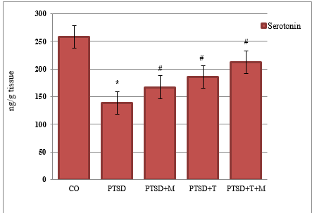
Figure 2. Comparison of Serotonin means in experiences and control groups
*: Significant difference with the control group
#: Significant difference with the PTSD group
Table5. Two-way ANOVA results for serotonin levels (ng/g tissue (
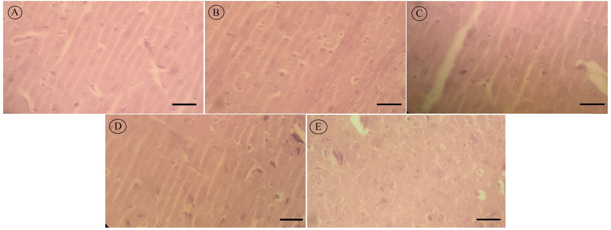
Figure 3. Comparison of H&E-stained hippocampal tissue samples across experimental groups: (A( HC group, (B) PTSD group, (C) PTSD+T group, (D) PTSD+M group, (E) PTSD+M+T group. In healthy controls, hippocampal neurons displayed normal morphology with high cellular density, round or oval nuclei, clear cytoplasmic borders, and a uniform matrix, with minimal abnormalities. In the PTSD group, neurons showed marked pathological changes, including shrinkage, nuclear condensation, apoptosis, and reduced cell density. Hippocampal tissue from PTSD rats subjected to combined exercise showed partial improvement, with scattered neurons, fewer dark nuclei, and signs of increased neurogenesis. Administration of milk thistle extract in PTSD rats resulted in a higher density of round, healthy neurons with larger nuclei and improved structural integrity. The combination of exercise and milk thistle extract produced the most pronounced protective effect, with dense, uniformly organized neurons closely resembling the healthy control group. Hematoxylin and eosin (H&E) staining ×400 magnification. Scale bar = 10 µm.
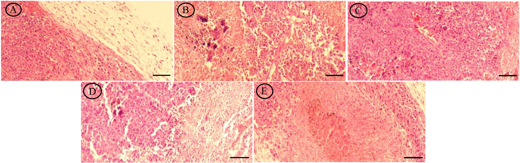
Figure 4. Comparison of H&E-stained liver tissue samples across experimental groups: (A( HC group, (B) PTSD group, (C) PTSD+T group, (D) PTSD+M group, (E) PTSD+M+T group. In healthy controls, hepatocytes were well-organized with regular, round nuclei, clear cytoplasmic borders, and uniform lobular architecture, showing minimal signs of cellular damage. In the PTSD group, hepatocytes exhibited marked pathological changes, including disorganization, nuclear condensation, cytoplasmic vacuolation, and reduced cell density. Liver tissue from PTSD rats subjected to combined exercise showed partial recovery, with improved hepatocyte arrangement and fewer abnormal nuclei. Administration of milk thistle extract in PTSD rats resulted in higher hepatocyte density, more regular nuclei, and restored lobular structure. The combination of exercise and milk thistle extract produced the most pronounced protective effect, with hepatocytes closely resembling the healthy control group, minimal vacuolation, and uniform tissue architecture. Hematoxylin and eosin (H&E) staining, ×400 magnification. Scale bar = 50 µm.
Farah Nameni *1
- Department of Exercise Physiology, VaP. C., Islamic Azad University, Varamin, Iran
- Science and Research Branch, Islamic Azad University, Tehran, Iran
Article history
Received 25 Mar 2026
Accepted 9 Jun 2026
A B S T R A C T
Introduction: Post-traumatic stress disorder (PTSD) is characterized by chronic neuroinflammation, impaired serotonergic signaling, and structural damage in both central and peripheral tissues, including the hippocampus and liver. Nuclear factor kappa B (NF-κB) plays a central role in stress-induced inflammatory responses. Exercise and phytotherapeutic agents such as milk thistle (Silybum marianum) have been suggested as potential non-pharmacological strategies for attenuating PTSD-related pathophysiology.
Methods: Fifty-five male Wistar rats were randomly assigned to five groups: healthy control, PTSD, PTSD with concurrent exercise training, PTSD with milk thistle extract, and PTSD with concurrent exercise training plus milk thistle extract. PTSD was induced using a multi-stage stress protocol. The concurrent exercise training program was conducted for 4 weeks, consisting of resistance training on even days and aerobic training on odd days. Resistance training was performed with progressively increasing loads from 50% to 80% of body weight, and aerobic training from 50% to 80% of VO₂max. Milk thistle extract was administered daily at a dose of 300 mg/kg. After the intervention, hippocampal and liver tissues were collected. NF-κB mRNA expression was measured using real-time PCR, and serotonin levels were measured by ELISA. Histopathological alterations were assessed by hematoxylin and eosin staining. Data were analyzed using ANOVA and Tukey statistical tests.
Results: PTSD induction significantly increased NF-κB mRNA expression and decreased serotonin levels in both hippocampal and liver tissues compared with healthy controls. Concurrent exercise training and milk thistle extract each significantly attenuated NF-κB overexpression and partially restored serotonin levels (p < 0.05). Histopathological analysis revealed severe neuronal degeneration in the hippocampus and marked structural damage in liver tissue in PTSD rats, whereas both interventions, particularly their combination, markedly improved tissue morphology.
Conclusion: Concurrent exercise training and milk thistle extract exert synergistic neuroprotective and hepatoprotective effects in a rat model of PTSD. These benefits appear to be mediated through suppression of NF-κB–dependent inflammatory pathways and restoration of serotonergic homeostasis.
Keywords: PTSD, Exercise Training, Milk Thistle Extract, Serotonin, NF-κB
Copyright © 2026 The Author(s). This is an open-access article distributed under the terms of the Creative Commons Attribution-Non Commercial 4.0 International License (http://creativecommons.org/licenses/by-nc/4.0/) which permits copy and redistribute the material just in noncommercial usages, provided the original work is properly cite.
Introduction
Post-Traumatic Stress Disorder (PTSD) is a chronic and complex stress-related psychiatric condition life-threatening. This disorder is characterized by persistent symptoms such as intrusive memories, avoidance behaviors, alterations, and heightened arousal. Epidemiological evidence indicates that PTSD not only significantly impairs quality of life but is also associated with an increased risk of depression, chronic anxiety, cognitive dysfunction, and neurodegenerative disorders (1). From a neurobiological perspective, PTSD is a multifactorial condition hypothalamic–pituitary–adrenal (HPA) axis immune responses, neurotransmitter systems, and molecular inflammatory pathways (2).
Neurobiological studies have demonstrated that prolonged exposure to severe stress induces significant structural and functional alterations in key brain regions, including the hippocampus, amygdala, and prefrontal cortex (3). These alterations involve reduced hippocampal volume, impaired synaptic plasticity, decreased neurogenesis, and increased neuronal apoptosis. At the molecular level, chronic stress activates inflammatory signaling pathways and enhances the production of reactive oxygen species, ultimately leading to neuronal damage and disruption of normal brain tissue architecture (4).
Among the neurotransmitter systems involved in the pathophysiology of PTSD, serotonin (5-hydroxytryptamine; 5-HT) plays a pivotal role. Serotonin is critically involved in the regulation of mood, anxiety, sleep, learning, and memory, and dysregulation of the serotonergic system has been strongly linked to the development of depressive- and anxiety-like behaviors in PTSD (5). Reduced serotonin levels and impaired receptor signaling contribute to increased stress sensitivity and diminished adaptive capacity of the brain. Consequently, many pharmacological treatments for PTSD primarily target the serotonergic system; however, these therapies often exhibit limited efficacy and undesirable side effects (6).
PTSD is a systemic stress-related condition that affects not only brain function but also peripheral organs, including the liver, via prolonged activation (7). Chronic stress associated with PTSD has been shown to induce hepatic oxidative stress, inflammatory responses, and structural alterations in liver tissue, largely mediated by dysregulation of the hypothalamic–pituitary–adrenal axis and activation of inflammatory signaling pathways such as nuclear factor kappa B (NF-κB) (8). Given the central role of the liver in metabolic homeostasis and detoxification, stress-induced hepatic damage may further exacerbate neuroinflammation and neurotransmitter imbalance via the gut–liver–brain axis. Concurrent exercise training has been reported to improve hepatic antioxidant capacity, attenuate inflammation, and preserve liver tissue integrity, while milk thistle (Silybum marianum) extract, rich in silymarin, is widely recognized for its hepatoprotective, anti-inflammatory, and antioxidant properties (9). Therefore, investigating the combined effects of exercise and milk thistle extract on liver tissue, alongside neurobiological markers, may provide a more comprehensive understanding of the systemic protective mechanisms underlying non-pharmacological interventions in PTSD (10). In addition to neurotransmitter dysregulation, neuroinflammation has emerged a central mechanism underlying PTSD-related brain pathology. Nuclear factor kappa B (NF-κB) is a key transcription factor regulating inflammatory responses within the central nervous system and is markedly activated under conditions of severe and chronic stress. Activation of NF-κB leads to increased expression of pro-inflammatory cytokines, inflammatory enzymes, and apoptosis-related factors (11). Experimental evidence from animal models indicates that excessive NF-κB activation is associated with enhanced neuroinflammation, reduced neuroplasticity, and impairment of serotonergic neurotransmission. Therefore, modulation of the NF-κB signaling pathway represents a promising therapeutic target for mitigating PTSD-induced neural damage (12).
In recent years, physical exercise has been widely recognized as an effective non-pharmacological intervention for improving brain health and alleviating psychological disorders. Concurrent exercise training, incorporating both aerobic and resistance components, exerts synergistic effects on multiple physiological and neural pathways (13). Evidence suggests that concurrent exercise training enhances cerebral blood flow, stimulates neurogenesis, improves synaptic plasticity, and increases the production of neurotransmitters such as serotonin. Moreover, regular physical activity has been shown to attenuate oxidative stress and suppress NF-κB activation, thereby reducing neuroinflammation and protecting brain tissue against stress-induced damage (14).
Alongside exercise interventions, increasing attention has been directed toward the neuroprotective potential of natural and herbal compounds. Milk thistle (Silybum marianum) extract, rich in bioactive flavonolignans such as silymarin, possesses potent antioxidant, anti-inflammatory, and neuroprotective properties (15). Experimental studies have demonstrated that silymarin can inhibit NF-κB activation, reduce oxidative stress, and enhance cellular antioxidant defenses, thereby preventing neuronal injury. Furthermore, emerging evidence suggests that milk thistle extract may positively modulate neurotransmitter balance, particularly within the serotonergic system, contributing to improvements in anxiety and depression-like behaviors (16).
Given the ethical and technical limitations of directly examining brain alterations in humans, animal models particularly male rats serve as valuable tools for investigating the underlying mechanisms of PTSD. Experimental PTSD models allow for controlled stress exposure, detailed assessment of molecular and biochemical alterations, and precise evaluation of brain tissue pathology. Measurement of inflammatory and neurochemical markers such as NF-κB and serotonin using immunoassay techniques, combined with histopathological analysis of brain tissue, provides a comprehensive framework for evaluating the effects of exercise and nutritional interventions.
Despite growing evidence supporting the individual benefits of exercise and herbal supplementation, limited studies have examined their combined effects on neuroinflammatory pathways, neurotransmitter regulation, and brain tissue integrity in PTSD. Therefore, the present study aims to investigate the role of concurrent exercise training and milk thistle extract consumption on brain tissue morphology, serotonin levels, and NF-κB-mediated inflammatory signaling in a rat model of PTSD. It is anticipated that the findings of this study will provide novel experimental evidence regarding the synergistic neuroprotective effects of exercise and herbal supplementation and may contribute to the development of complementary, low-risk therapeutic strategies for PTSD.
Methods
Study design and procedures
The statistical population included 55 Wistar rats (11 rats per group), aged 14-16 weeks and weighing 280–320 g, which were obtained from the Laboratory Animal Breeding Center of the Pasteur Institute and transferred to the university animal facility. Before the intervention, a one-week adaptation period was allowed under standard laboratory conditions: a 12:12 h light/dark cycle, temperature of 22 ± 3°C, and relative humidity of 50–60%. During this time, the animals had free access to standard pellet food and water.
Sample size
The inclusion criteria were healthy male Wistar rats, 14–16 weeks old, weighing 280–320 g, no history of disease or drug treatment, and at least one week of environmental adaptation. Exclusion criteria included the appearance of disease symptoms or abnormal behaviors, body weight loss greater than 20% during the study, failure to comply with the training protocol, or death during the experimental period. The animals were housed in polycarbonate cages, five rats per cage, under standard laboratory conditions. Based on a pilot study, a power of 80% and an alpha of 0.05 were considered.
Induction of the PTSD model
The PTSD model in rats was induced using a multi-stage protocol combining physical restraint, forced swimming, and chemical stress exposure. In the first stage, animals were restrained for 2 hours in a 100-mL cylindrical tube (4cm diameter) to induce complete immobilization, representing acute physical and psychological stress. In the second stage, immediately following restraint, rats were subjected to forced swimming in an animal pool for 15 minutes to elicit active stress responses. In the third stage, after a 15-minute rest, animals were exposed to an ether-filled chamber for 3 minutes, inducing transient anesthesia and additional chemical stress (17). This multimodal stress protocol effectively models the physical, psychological, and chemical stressors associated with PTSD in rats. This multimodal stress provides a controlled and reproducible approach to study the systemic effects of stress on cardiac tissue and gene/protein expression, allowing the investigation of potential protective interventions such as Silybum marianum.
Following the induction of combined stress, the animals underwent behavioral assessments 7 days after exposure. Only animals that met at least two independent behavioral criteria defined as values exceeding the mean ± 2 standard deviations of the control group were deemed eligible for inclusion in the subsequent phases of the study.
In the Open Field Test (OFT), animals classified as PTSD‑like were required to show less than 10 seconds spent in the central zone (control:24.6 ± 7.2s) and less than 5% of the total distance traveled in the central zone (control:9.8 ± 2.4%). For the Elevated Plus Maze (EPM), control animals exhibited 22.4 ± 6.1% time spent in the open arms and 8.1 ± 1.9 open‑arm entries. Therefore, in our study, animals were categorized as having a valid PTSD phenotype only if they spent less than 10% of the time in the open arms and made fewer than 4 open‑arm entries.
In the startle response test, animals displaying a startle amplitude greater than 220 units (control: 148 ± 36) and a response latency greater than 300 ms (control: 21.3 ± 4.7 ms) were classified as having a valid PTSD phenotype for this behavioral domain. Animals that met these criteria were included, and the remaining animals were excluded from further analyses.
Following the PTSD protocol, rats were randomly assigned to five groups: 1) healthy control group (C); 2) PTSD group (P), which received stress only; 3) exercise group (E), which received the training protocol in addition to stress; 4) milk thistle group (S), which received milk thistle in addition to stress; and 5) combined group (E+S), which received both interventions. The therapeutic interventions began 14 days after PTSD induction, allowing time for the symptoms to stabilize (18).
To minimize potential bias, animals were randomly assigned to experimental groups using a random number table by an independent researcher who was not involved in other parts of the study. Randomization was validated by confirming that baseline body weights did not differ significantly among groups. Behavioral and tissue assessments were performed by an investigator blinded to group assignments to ensure objectivity and reduce assessment bias.
To ensure accurate to validate of the PTSD-like phenotype, to exclude non-responders, and to confirm PTSD induction, animals were required to perform three tests: Open Field Test (OFT), Elevated plus Maze (EPM), and Startle Response Test.
Evaluation and validation of the PTSD model
To complement this assessment, the startle response test was performed, which measures the intensity of the startle reflex in response to a sudden acoustic stimulus and serves as a sensitive index for hyper arousal, hyper vigilance, and fear reactivation in PTSD. Using these stringent criteria, 65.4% of SPS-exposed rats (29 of 44) met the classification requirements and were included in subsequent molecular and biochemical analyses. To accurately validate the PTSD-like phenotype, exclude non-responders, and confirm PTSD induction, animals were required to perform three tests with values that were greater than the mean ± 2 standard deviations (SD) of the unstressed control group .In the Open Field Test (5-min session in a 50×50 cm arena), animals classified as PTSD showed the following:(1) central zone time < 10s (control mean ± SD: 24.6 ± 7.2s) and (2) percentage of distance traveled in the central zone < 5% (control: 9.8 ± 2.4%), while total distance traveled did not differ significantly between groups (control: 2845 ± 218 cm; classified as PTSD: 2693 ± 241 cm, p > 0.05), confirming that the decreased central exploration reflects increased anxiety-like behavior rather than reduced locomotion. Animals failing to meet both OFT criteria (and/or the second behavioral domain criteria) were excluded to maintain phenotypic homogeneity, in accordance with current best practice recommendations for preclinical PTSD research (19). The observation of consistent patterns across the tests-namely, high anxiety in the EPM and exaggerated startle response confirms that the PTSD model used in this study exhibited sufficient behavioral validity.
Combined training protocol
Fourteen days after PTSD induction, the combined training protocol was initiated, consisting of combining resistance and aerobic training. All training sessions were performed using a specialized animal treadmill (Tajhiz Gostar Iranian, Model 2020) and a custom-built vertical ladder. The training was structured to include three resistance sessions and three aerobic sessions per week, conducted on alternating days. Resistance training was carried out on Saturdays, Mondays, and Wednesdays. Each session consisted of three sets, with each set comprising four ascents of a 1-meter-high vertical ladder with 26 steps (4 cm apart). A 30-second rest was provided between each climb. To apply resistance, a weight was attached to the animals' tails, and tail stimulation was used to encourage continuous movement. The principle of progressive overload was implemented by increasing the percentage of body weight carried by the animals every week. Specifically, the weight was 50% of the animal's body weight in the first week, 60% in the second week, 70% in the third week, and 80% in the fourth week. Aerobic training sessions were performed on Sundays, Tuesdays, and Thursdays for a total of 4 weeks. Before the main training program, the animals were exposed to a 3-day familiarization program with treadmill running. Following this period, each rat's maximal oxygen consumption (VO2max) was determined using a graded exercise protocol. The average of these records was then used as a reference to design the main training program in terms of both distance and time, ensuring that the intensity was tailored to each animal's capacity (20). Fatigue was assessed using a combination of behavioral and physiological criteria to ensure maximal exertion without undue stress to the animals. Before implementing the main protocol, a pilot study was conducted with several rats to establish normative values. Physiologically, an increase in respiratory exchange ratio (RER) to > 1.05 indicating a shift to anaerobic metabolism which typically occurred during the final stages of the incremental VO2max test on the treadmill (5°incline). Specifically, following a 15-min warm-up at 40–50% VO2max (speed ≈10–12 m/min), with speed increments of 1.8 m/min every 2 min, the RER threshold of 1.05 was reached at a mean time of 18 ± 3 min and speed of 26 ± 2 m/min (equivalent to 75–85%Vmax ≈35 m/min). These values aligned with the VO₂ plateau and elevated blood lactate levels and were derived from measurements in 8 rats using indirect calorimetry. Behaviorally, fatigue was also indicated if the animal failed to maintain treadmill speed despite gentle tail stimulation, defined as falling off the belt more than 3–5 consecutive times or contacting the shock grid at the belt's end for more than 5 seconds. Running speeds were calculated based on each rat’s individual Vmax obtained from the VO2max test, and all training sessions were performed at a constant treadmill incline of 5%.
Preparation of milk thistle extract
Milk thistle seeds was stored in sealed containers at low temperature to preserve bioactive compounds. One hundred grams of powdered seeds were used for extraction. Defatting was carried out to remove nonpolar lipids. The defatted residue was extracted with absolute ethanol (96%,10mL/g). The combined extracts were concentrated to yield a yellowish-brown powder. The extract was stored at -20°C until use. Silymarin content was quantified by high-performance liquid chromatography (HPLC). Detection was performed at 285 nm with a total runtime of 16.5 min. For analysis, the dried extract was dissolved in methanol (1mg/mL), diluted with 50% aqueous methanol, and filtered through a membrane. Calibration curves were prepared using authenticated standards and were validated. The initial extract (~50% silymarin) was standardized to 30% by blending with microcrystalline cellulose, and HPLC analysis confirmed 300mg silymarin per gram of dry extract. The standardized extract remained stable for six months at -80 °C. Quality control included thin-layer chromatography (TLC). The extract was administered orally to rats via gavage at 300mg/kg body weight, a dose selected based on previous studies demonstrating maximal hepatoprotective and antioxidant effects (21). The vehicle consisted of 0.5% Polysorbate 80 dissolved in sterile distilled water. The extract was dissolved/suspended in the aforementioned vehicle to achieve the desired dose within a final gavage volume of 5 mL/kg.
Administration of milk thistle via oral gavage
The prepared ethanolic extract of milk thistle was administered to the rats in the milk thistle and combined treatment groups oral gavage. This method was chosen to ensure the precise and controlled delivery of the substance directly into the stomach, thereby bypassing any potential degradation or dose-related effects that could occur in the oral cavity or during early digestive processes.
For daily administration, the required dose of milk thistle was calculated for each rat at 300 mg/kg of its most recent body weight. The calculated amount of the powdered extract was then freshly dissolved in an appropriate vehicle, which could be sterile distilled water or a saline solution. To improve the solubility of milk thistle and ensure a uniform suspension, a small amount of a wetting agent, such as Tween 80, was added to the solution. The prepared solution was administered daily at a consistent time to all animals in the respective treatment groups throughout the study period.
The gavage procedure was performed meticulously to ensure accuracy and safety. Each rat was carefully restrained, and a specialized gavage needle (a curved needle with a blunt, rounded tip) was gently inserted into the mouth and navigated down the esophagus, taking care to avoid the trachea. Once the correct placement of the needle was confirmed, the solution was slowly and steadily infused into the stomach. This precise and systematic procedure guaranteed that each animal received its exact, predetermined dose, which was critical for ensuring the reliability and reproducibility of the experimental results.
Euthanasia & anesthesia
After Forty-eight hours from the last training session, at the end of the 4-week intervention, the rats were deeply anesthetized with an intraperitoneal injection of ketamine (80 mg/kg) and xylazine (10 mg/kg). Deep anesthesia was confirmed by the loss of reflexes, followed by cervical dislocation to ensure euthanasia. All procedures were conducted in accordance with the institutional guidelines for the care and use of laboratory animals and were approved by the Institutional Animal Ethics Committee.
Tissue dissection
Hippocampal tissue was collected immediately after euthanasia. The brain was rapidly removed and placed on ice, and the hippocampus was carefully dissected under a stereomicroscope. The tissue was snap-frozen in liquid nitrogen and stored at -80°C until analysis. For ELISA, the hippocampal samples were later homogenized and processed according to the kit protocol to measure neurotransmitters, NF-κB, and serotonin. Low temperature was maintained, and handling time was minimized to preserve protein stability and ensure accurate results.
Liver tissue was collected immediately after euthanasia. The abdominal cavity was opened, and the liver was carefully removed, rinsed with ice-cold phosphate-buffered saline to remove blood, and placed on ice. The tissue was then snap-frozen in liquid nitrogen and stored at -80°C until analysis. For ELISA, the liver samples were later homogenized and processed according to the kit protocol. Low temperature was maintained, and handling time was minimized to preserve protein stability and ensure accurate results.
Hippocampal and liver tissues were collected as previously described, immediately snap-frozen in liquid nitrogen, and stored at -80°C until analysis. Primers for NF-κB were designed using Primer3 software (version 2.6.1) and synthesized with HPLC-grade purity. Primer sequences and amplicon sizes were optimized for qRT-PCR using SYBR Green Master Mix, with amplification efficiencies ranging from 90–110%. Primer specificity was confirmed by melt curve analysis and amplicon sequencing. This setup allowed accurate measurement of NF-κB mRNA expression in hippocampal and liver tissues of male rats in the PTSD model.
Serotonin measurement by ELISA
Hippocampal tissues were collected, snap frozen in liquid nitrogen, and stored at –80°C until analysis. Before assay, tissues were thawed on ice, weighed, and homogenized in ice-cold phosphate-buffered saline containing protease inhibitors. Homogenates were centrifuged at 10,000×g for 10 minutes at 4°C, and the supernatants were collected for analysis. Serotonin concentrations were measured using a commercial ELISA kit according to the manufacturer’s instructions. All samples and standards were assayed in triplicate to ensure accuracy and reproducibility. Absorbance was read at the specified wavelength using a microplate reader, and serotonin levels were calculated using standard curves. Final concentrations were expressed in nanograms per gram of tissue (ng/g tissue). Low temperature was maintained throughout all procedures, and freeze-thaw cycles were minimized to preserve neurotransmitter integrity (My BioSource Catalog No. MBS166089; detection range 0.5–200 ng/mL, sensitivity 0.23 ng/mL). Absorbance was measured at 450 nm using a micro plate reader and serotonin concentrations were calculated from standard curves.
Histological imaging and analysis
Representative images of hippocampal and liver sections were captured following H&E staining using a light microscope equipped with a digital camera. For each tissue type, multiple fields of view were examined at different magnifications to ensure consistency and representative sampling. The images were used to qualitatively compare tissue morphology, cellular density, and pathological changes among experimental groups. Quantitative analysis was performed where applicable, such as measuring hepatocyte size, nuclear-to-cytoplasmic ratio, or counting degenerative cells in the hippocampus. ImageJ software (Fiji version 2.1.0, NIH, USA) used to standardize measurements. All images were processed under identical brightness and contrast settings to allow direct comparison between groups.
Statistical analysis
Data analysis was performed using SPSS version 19. First, the normality of the data distribution was assessed using the Shapiro-Wilk test, and homogeneity of variances was evaluated with the Levene test. Descriptive statistics were presented as mean ± standard deviation (SD). Between-group differences were analyzed using one-way ANOVA. Tukey post hoc test was applied to determine the specific location of significant differences (p < 0.05). Two-way methods were used to examine the effect of each intervention (p < 0.05). Effect sizes were calculated using Cohen’s d for pairwise comparisons, and Eta squared (η²) for ANOVA to quantify the magnitude of observed effects.
Ethical considerations
All procedures were approved by the Institutional Animal Care and Use Committee and reported in accordance with the ARRIVE guidelines and the study was approved by the Science and Research Branch, Islamic Azad University Ethics Committee (Ethics code: IR.IAU.SRB.REC.1404.199) and carried out in accordance with international ethical guidelines for the care and use of laboratory animals
Results
Validation of the PTSD model
Open Field Test (OFT) results demonstrated that rats exposed to the PTSD protocol exhibited a significant reduction in central area exploration, as reflected by decreased central time and central distance compared with the control group, while total locomotor activity did not differ significantly among groups, indicating anxiety-like behavior rather than impaired locomotion. Both exercise and milk thistle extract partially improved central exploration, and rats receiving the combined intervention showed the greatest improvement, with central exploration approaching control levels.
Behavioral validation of PTSD induction
To verify that PTSD was successfully induced before any therapeutic intervention, all animals underwent a two-stage behavioral and hormonal assessment including baseline (pre-induction) and post-induction evaluations. The baseline testing battery included the Open Field Test (OFT), Elevated plus Maze (EPM), and Acoustic Startle Response. Blood sampling was also
performed before and after induction to evaluate HPA axis activity through serum cortisol levels. All assessments were carried out at the same time window (09:00–11:00) to minimize circadian effects.
Body weight changes
The body weights of rats were monitored the study across the five experimental groups: control, PTSD, combined training, milk thistle extract, and combined training with milk thistle extract. Descriptive analysis (Mean ± SD and paired t-tests) indicated that all groups began with comparable baseline weights (p > 0.05). (Table1)
A slight but not statistically significant reduction in body weight was observed. In contrast, rats in the training-only and extract-only groups generally maintained or slightly increased their weights. The combined training with milk thistle extract group demonstrated a modest but significant increase in body weight compared with the PTSD group (p < 0.05). Two-way ANOVA revealed significant main effects of both intervention type and PTSD condition on body weight (p < 0.05). Post hoc analysis (Tukey test) showed that the combined intervention group had a higher mean body weight than the PTSD-only group, whereas no significant differences were detected between the control and single-intervention groups. Weight gain in treated groups likely reflects improved appetite and reduced stress. The results are statistically significant (p < 0.03), but caution should be exercised in interpretation due to the relatively small size of the groups. The results showed that before the intervention, there was no significant difference between the values of the dependent variables in the two groups of healthy and PTSD-induced rats.
The changes in NF-κB gene expression and serotonin between groups were analyzed by one-way ANOVA. (Figure 1, Table 2)
One-way ANOVA revealed a significant increase in NF-κB mRNA expression in the groups. Tukey's post hoc test showed a significant difference between the means among the 5 experimental and control groups. (Table3)
To evaluate the effects of combined exercise training and milk thistle extract on NF-κB mRNA expression, a two-way ANOVA was conducted with exercise and supplementation as independent variables. NF-κB mRNA expression was considered the dependent variable, and effect sizes were calculated using partial eta squared (η²). (Table 4)
Neurobiological studies have demonstrated that prolonged exposure to severe stress induces significant structural and functional alterations in key brain regions, including the hippocampus, amygdala, and prefrontal cortex (3). These alterations involve reduced hippocampal volume, impaired synaptic plasticity, decreased neurogenesis, and increased neuronal apoptosis. At the molecular level, chronic stress activates inflammatory signaling pathways and enhances the production of reactive oxygen species, ultimately leading to neuronal damage and disruption of normal brain tissue architecture (4).
Among the neurotransmitter systems involved in the pathophysiology of PTSD, serotonin (5-hydroxytryptamine; 5-HT) plays a pivotal role. Serotonin is critically involved in the regulation of mood, anxiety, sleep, learning, and memory, and dysregulation of the serotonergic system has been strongly linked to the development of depressive- and anxiety-like behaviors in PTSD (5). Reduced serotonin levels and impaired receptor signaling contribute to increased stress sensitivity and diminished adaptive capacity of the brain. Consequently, many pharmacological treatments for PTSD primarily target the serotonergic system; however, these therapies often exhibit limited efficacy and undesirable side effects (6).
PTSD is a systemic stress-related condition that affects not only brain function but also peripheral organs, including the liver, via prolonged activation (7). Chronic stress associated with PTSD has been shown to induce hepatic oxidative stress, inflammatory responses, and structural alterations in liver tissue, largely mediated by dysregulation of the hypothalamic–pituitary–adrenal axis and activation of inflammatory signaling pathways such as nuclear factor kappa B (NF-κB) (8). Given the central role of the liver in metabolic homeostasis and detoxification, stress-induced hepatic damage may further exacerbate neuroinflammation and neurotransmitter imbalance via the gut–liver–brain axis. Concurrent exercise training has been reported to improve hepatic antioxidant capacity, attenuate inflammation, and preserve liver tissue integrity, while milk thistle (Silybum marianum) extract, rich in silymarin, is widely recognized for its hepatoprotective, anti-inflammatory, and antioxidant properties (9). Therefore, investigating the combined effects of exercise and milk thistle extract on liver tissue, alongside neurobiological markers, may provide a more comprehensive understanding of the systemic protective mechanisms underlying non-pharmacological interventions in PTSD (10). In addition to neurotransmitter dysregulation, neuroinflammation has emerged a central mechanism underlying PTSD-related brain pathology. Nuclear factor kappa B (NF-κB) is a key transcription factor regulating inflammatory responses within the central nervous system and is markedly activated under conditions of severe and chronic stress. Activation of NF-κB leads to increased expression of pro-inflammatory cytokines, inflammatory enzymes, and apoptosis-related factors (11). Experimental evidence from animal models indicates that excessive NF-κB activation is associated with enhanced neuroinflammation, reduced neuroplasticity, and impairment of serotonergic neurotransmission. Therefore, modulation of the NF-κB signaling pathway represents a promising therapeutic target for mitigating PTSD-induced neural damage (12).
In recent years, physical exercise has been widely recognized as an effective non-pharmacological intervention for improving brain health and alleviating psychological disorders. Concurrent exercise training, incorporating both aerobic and resistance components, exerts synergistic effects on multiple physiological and neural pathways (13). Evidence suggests that concurrent exercise training enhances cerebral blood flow, stimulates neurogenesis, improves synaptic plasticity, and increases the production of neurotransmitters such as serotonin. Moreover, regular physical activity has been shown to attenuate oxidative stress and suppress NF-κB activation, thereby reducing neuroinflammation and protecting brain tissue against stress-induced damage (14).
Alongside exercise interventions, increasing attention has been directed toward the neuroprotective potential of natural and herbal compounds. Milk thistle (Silybum marianum) extract, rich in bioactive flavonolignans such as silymarin, possesses potent antioxidant, anti-inflammatory, and neuroprotective properties (15). Experimental studies have demonstrated that silymarin can inhibit NF-κB activation, reduce oxidative stress, and enhance cellular antioxidant defenses, thereby preventing neuronal injury. Furthermore, emerging evidence suggests that milk thistle extract may positively modulate neurotransmitter balance, particularly within the serotonergic system, contributing to improvements in anxiety and depression-like behaviors (16).
Given the ethical and technical limitations of directly examining brain alterations in humans, animal models particularly male rats serve as valuable tools for investigating the underlying mechanisms of PTSD. Experimental PTSD models allow for controlled stress exposure, detailed assessment of molecular and biochemical alterations, and precise evaluation of brain tissue pathology. Measurement of inflammatory and neurochemical markers such as NF-κB and serotonin using immunoassay techniques, combined with histopathological analysis of brain tissue, provides a comprehensive framework for evaluating the effects of exercise and nutritional interventions.
Despite growing evidence supporting the individual benefits of exercise and herbal supplementation, limited studies have examined their combined effects on neuroinflammatory pathways, neurotransmitter regulation, and brain tissue integrity in PTSD. Therefore, the present study aims to investigate the role of concurrent exercise training and milk thistle extract consumption on brain tissue morphology, serotonin levels, and NF-κB-mediated inflammatory signaling in a rat model of PTSD. It is anticipated that the findings of this study will provide novel experimental evidence regarding the synergistic neuroprotective effects of exercise and herbal supplementation and may contribute to the development of complementary, low-risk therapeutic strategies for PTSD.
Methods
Study design and procedures
The statistical population included 55 Wistar rats (11 rats per group), aged 14-16 weeks and weighing 280–320 g, which were obtained from the Laboratory Animal Breeding Center of the Pasteur Institute and transferred to the university animal facility. Before the intervention, a one-week adaptation period was allowed under standard laboratory conditions: a 12:12 h light/dark cycle, temperature of 22 ± 3°C, and relative humidity of 50–60%. During this time, the animals had free access to standard pellet food and water.
Sample size
The inclusion criteria were healthy male Wistar rats, 14–16 weeks old, weighing 280–320 g, no history of disease or drug treatment, and at least one week of environmental adaptation. Exclusion criteria included the appearance of disease symptoms or abnormal behaviors, body weight loss greater than 20% during the study, failure to comply with the training protocol, or death during the experimental period. The animals were housed in polycarbonate cages, five rats per cage, under standard laboratory conditions. Based on a pilot study, a power of 80% and an alpha of 0.05 were considered.
Induction of the PTSD model
The PTSD model in rats was induced using a multi-stage protocol combining physical restraint, forced swimming, and chemical stress exposure. In the first stage, animals were restrained for 2 hours in a 100-mL cylindrical tube (4cm diameter) to induce complete immobilization, representing acute physical and psychological stress. In the second stage, immediately following restraint, rats were subjected to forced swimming in an animal pool for 15 minutes to elicit active stress responses. In the third stage, after a 15-minute rest, animals were exposed to an ether-filled chamber for 3 minutes, inducing transient anesthesia and additional chemical stress (17). This multimodal stress protocol effectively models the physical, psychological, and chemical stressors associated with PTSD in rats. This multimodal stress provides a controlled and reproducible approach to study the systemic effects of stress on cardiac tissue and gene/protein expression, allowing the investigation of potential protective interventions such as Silybum marianum.
Following the induction of combined stress, the animals underwent behavioral assessments 7 days after exposure. Only animals that met at least two independent behavioral criteria defined as values exceeding the mean ± 2 standard deviations of the control group were deemed eligible for inclusion in the subsequent phases of the study.
In the Open Field Test (OFT), animals classified as PTSD‑like were required to show less than 10 seconds spent in the central zone (control:24.6 ± 7.2s) and less than 5% of the total distance traveled in the central zone (control:9.8 ± 2.4%). For the Elevated Plus Maze (EPM), control animals exhibited 22.4 ± 6.1% time spent in the open arms and 8.1 ± 1.9 open‑arm entries. Therefore, in our study, animals were categorized as having a valid PTSD phenotype only if they spent less than 10% of the time in the open arms and made fewer than 4 open‑arm entries.
In the startle response test, animals displaying a startle amplitude greater than 220 units (control: 148 ± 36) and a response latency greater than 300 ms (control: 21.3 ± 4.7 ms) were classified as having a valid PTSD phenotype for this behavioral domain. Animals that met these criteria were included, and the remaining animals were excluded from further analyses.
Following the PTSD protocol, rats were randomly assigned to five groups: 1) healthy control group (C); 2) PTSD group (P), which received stress only; 3) exercise group (E), which received the training protocol in addition to stress; 4) milk thistle group (S), which received milk thistle in addition to stress; and 5) combined group (E+S), which received both interventions. The therapeutic interventions began 14 days after PTSD induction, allowing time for the symptoms to stabilize (18).
To minimize potential bias, animals were randomly assigned to experimental groups using a random number table by an independent researcher who was not involved in other parts of the study. Randomization was validated by confirming that baseline body weights did not differ significantly among groups. Behavioral and tissue assessments were performed by an investigator blinded to group assignments to ensure objectivity and reduce assessment bias.
To ensure accurate to validate of the PTSD-like phenotype, to exclude non-responders, and to confirm PTSD induction, animals were required to perform three tests: Open Field Test (OFT), Elevated plus Maze (EPM), and Startle Response Test.
Evaluation and validation of the PTSD model
To complement this assessment, the startle response test was performed, which measures the intensity of the startle reflex in response to a sudden acoustic stimulus and serves as a sensitive index for hyper arousal, hyper vigilance, and fear reactivation in PTSD. Using these stringent criteria, 65.4% of SPS-exposed rats (29 of 44) met the classification requirements and were included in subsequent molecular and biochemical analyses. To accurately validate the PTSD-like phenotype, exclude non-responders, and confirm PTSD induction, animals were required to perform three tests with values that were greater than the mean ± 2 standard deviations (SD) of the unstressed control group .In the Open Field Test (5-min session in a 50×50 cm arena), animals classified as PTSD showed the following:(1) central zone time < 10s (control mean ± SD: 24.6 ± 7.2s) and (2) percentage of distance traveled in the central zone < 5% (control: 9.8 ± 2.4%), while total distance traveled did not differ significantly between groups (control: 2845 ± 218 cm; classified as PTSD: 2693 ± 241 cm, p > 0.05), confirming that the decreased central exploration reflects increased anxiety-like behavior rather than reduced locomotion. Animals failing to meet both OFT criteria (and/or the second behavioral domain criteria) were excluded to maintain phenotypic homogeneity, in accordance with current best practice recommendations for preclinical PTSD research (19). The observation of consistent patterns across the tests-namely, high anxiety in the EPM and exaggerated startle response confirms that the PTSD model used in this study exhibited sufficient behavioral validity.
Combined training protocol
Fourteen days after PTSD induction, the combined training protocol was initiated, consisting of combining resistance and aerobic training. All training sessions were performed using a specialized animal treadmill (Tajhiz Gostar Iranian, Model 2020) and a custom-built vertical ladder. The training was structured to include three resistance sessions and three aerobic sessions per week, conducted on alternating days. Resistance training was carried out on Saturdays, Mondays, and Wednesdays. Each session consisted of three sets, with each set comprising four ascents of a 1-meter-high vertical ladder with 26 steps (4 cm apart). A 30-second rest was provided between each climb. To apply resistance, a weight was attached to the animals' tails, and tail stimulation was used to encourage continuous movement. The principle of progressive overload was implemented by increasing the percentage of body weight carried by the animals every week. Specifically, the weight was 50% of the animal's body weight in the first week, 60% in the second week, 70% in the third week, and 80% in the fourth week. Aerobic training sessions were performed on Sundays, Tuesdays, and Thursdays for a total of 4 weeks. Before the main training program, the animals were exposed to a 3-day familiarization program with treadmill running. Following this period, each rat's maximal oxygen consumption (VO2max) was determined using a graded exercise protocol. The average of these records was then used as a reference to design the main training program in terms of both distance and time, ensuring that the intensity was tailored to each animal's capacity (20). Fatigue was assessed using a combination of behavioral and physiological criteria to ensure maximal exertion without undue stress to the animals. Before implementing the main protocol, a pilot study was conducted with several rats to establish normative values. Physiologically, an increase in respiratory exchange ratio (RER) to > 1.05 indicating a shift to anaerobic metabolism which typically occurred during the final stages of the incremental VO2max test on the treadmill (5°incline). Specifically, following a 15-min warm-up at 40–50% VO2max (speed ≈10–12 m/min), with speed increments of 1.8 m/min every 2 min, the RER threshold of 1.05 was reached at a mean time of 18 ± 3 min and speed of 26 ± 2 m/min (equivalent to 75–85%Vmax ≈35 m/min). These values aligned with the VO₂ plateau and elevated blood lactate levels and were derived from measurements in 8 rats using indirect calorimetry. Behaviorally, fatigue was also indicated if the animal failed to maintain treadmill speed despite gentle tail stimulation, defined as falling off the belt more than 3–5 consecutive times or contacting the shock grid at the belt's end for more than 5 seconds. Running speeds were calculated based on each rat’s individual Vmax obtained from the VO2max test, and all training sessions were performed at a constant treadmill incline of 5%.
Preparation of milk thistle extract
Milk thistle seeds was stored in sealed containers at low temperature to preserve bioactive compounds. One hundred grams of powdered seeds were used for extraction. Defatting was carried out to remove nonpolar lipids. The defatted residue was extracted with absolute ethanol (96%,10mL/g). The combined extracts were concentrated to yield a yellowish-brown powder. The extract was stored at -20°C until use. Silymarin content was quantified by high-performance liquid chromatography (HPLC). Detection was performed at 285 nm with a total runtime of 16.5 min. For analysis, the dried extract was dissolved in methanol (1mg/mL), diluted with 50% aqueous methanol, and filtered through a membrane. Calibration curves were prepared using authenticated standards and were validated. The initial extract (~50% silymarin) was standardized to 30% by blending with microcrystalline cellulose, and HPLC analysis confirmed 300mg silymarin per gram of dry extract. The standardized extract remained stable for six months at -80 °C. Quality control included thin-layer chromatography (TLC). The extract was administered orally to rats via gavage at 300mg/kg body weight, a dose selected based on previous studies demonstrating maximal hepatoprotective and antioxidant effects (21). The vehicle consisted of 0.5% Polysorbate 80 dissolved in sterile distilled water. The extract was dissolved/suspended in the aforementioned vehicle to achieve the desired dose within a final gavage volume of 5 mL/kg.
Administration of milk thistle via oral gavage
The prepared ethanolic extract of milk thistle was administered to the rats in the milk thistle and combined treatment groups oral gavage. This method was chosen to ensure the precise and controlled delivery of the substance directly into the stomach, thereby bypassing any potential degradation or dose-related effects that could occur in the oral cavity or during early digestive processes.
For daily administration, the required dose of milk thistle was calculated for each rat at 300 mg/kg of its most recent body weight. The calculated amount of the powdered extract was then freshly dissolved in an appropriate vehicle, which could be sterile distilled water or a saline solution. To improve the solubility of milk thistle and ensure a uniform suspension, a small amount of a wetting agent, such as Tween 80, was added to the solution. The prepared solution was administered daily at a consistent time to all animals in the respective treatment groups throughout the study period.
The gavage procedure was performed meticulously to ensure accuracy and safety. Each rat was carefully restrained, and a specialized gavage needle (a curved needle with a blunt, rounded tip) was gently inserted into the mouth and navigated down the esophagus, taking care to avoid the trachea. Once the correct placement of the needle was confirmed, the solution was slowly and steadily infused into the stomach. This precise and systematic procedure guaranteed that each animal received its exact, predetermined dose, which was critical for ensuring the reliability and reproducibility of the experimental results.
Euthanasia & anesthesia
After Forty-eight hours from the last training session, at the end of the 4-week intervention, the rats were deeply anesthetized with an intraperitoneal injection of ketamine (80 mg/kg) and xylazine (10 mg/kg). Deep anesthesia was confirmed by the loss of reflexes, followed by cervical dislocation to ensure euthanasia. All procedures were conducted in accordance with the institutional guidelines for the care and use of laboratory animals and were approved by the Institutional Animal Ethics Committee.
Tissue dissection
Hippocampal tissue was collected immediately after euthanasia. The brain was rapidly removed and placed on ice, and the hippocampus was carefully dissected under a stereomicroscope. The tissue was snap-frozen in liquid nitrogen and stored at -80°C until analysis. For ELISA, the hippocampal samples were later homogenized and processed according to the kit protocol to measure neurotransmitters, NF-κB, and serotonin. Low temperature was maintained, and handling time was minimized to preserve protein stability and ensure accurate results.
Liver tissue was collected immediately after euthanasia. The abdominal cavity was opened, and the liver was carefully removed, rinsed with ice-cold phosphate-buffered saline to remove blood, and placed on ice. The tissue was then snap-frozen in liquid nitrogen and stored at -80°C until analysis. For ELISA, the liver samples were later homogenized and processed according to the kit protocol. Low temperature was maintained, and handling time was minimized to preserve protein stability and ensure accurate results.
Hippocampal and liver tissues were collected as previously described, immediately snap-frozen in liquid nitrogen, and stored at -80°C until analysis. Primers for NF-κB were designed using Primer3 software (version 2.6.1) and synthesized with HPLC-grade purity. Primer sequences and amplicon sizes were optimized for qRT-PCR using SYBR Green Master Mix, with amplification efficiencies ranging from 90–110%. Primer specificity was confirmed by melt curve analysis and amplicon sequencing. This setup allowed accurate measurement of NF-κB mRNA expression in hippocampal and liver tissues of male rats in the PTSD model.
Serotonin measurement by ELISA
Hippocampal tissues were collected, snap frozen in liquid nitrogen, and stored at –80°C until analysis. Before assay, tissues were thawed on ice, weighed, and homogenized in ice-cold phosphate-buffered saline containing protease inhibitors. Homogenates were centrifuged at 10,000×g for 10 minutes at 4°C, and the supernatants were collected for analysis. Serotonin concentrations were measured using a commercial ELISA kit according to the manufacturer’s instructions. All samples and standards were assayed in triplicate to ensure accuracy and reproducibility. Absorbance was read at the specified wavelength using a microplate reader, and serotonin levels were calculated using standard curves. Final concentrations were expressed in nanograms per gram of tissue (ng/g tissue). Low temperature was maintained throughout all procedures, and freeze-thaw cycles were minimized to preserve neurotransmitter integrity (My BioSource Catalog No. MBS166089; detection range 0.5–200 ng/mL, sensitivity 0.23 ng/mL). Absorbance was measured at 450 nm using a micro plate reader and serotonin concentrations were calculated from standard curves.
Histological imaging and analysis
Representative images of hippocampal and liver sections were captured following H&E staining using a light microscope equipped with a digital camera. For each tissue type, multiple fields of view were examined at different magnifications to ensure consistency and representative sampling. The images were used to qualitatively compare tissue morphology, cellular density, and pathological changes among experimental groups. Quantitative analysis was performed where applicable, such as measuring hepatocyte size, nuclear-to-cytoplasmic ratio, or counting degenerative cells in the hippocampus. ImageJ software (Fiji version 2.1.0, NIH, USA) used to standardize measurements. All images were processed under identical brightness and contrast settings to allow direct comparison between groups.
Statistical analysis
Data analysis was performed using SPSS version 19. First, the normality of the data distribution was assessed using the Shapiro-Wilk test, and homogeneity of variances was evaluated with the Levene test. Descriptive statistics were presented as mean ± standard deviation (SD). Between-group differences were analyzed using one-way ANOVA. Tukey post hoc test was applied to determine the specific location of significant differences (p < 0.05). Two-way methods were used to examine the effect of each intervention (p < 0.05). Effect sizes were calculated using Cohen’s d for pairwise comparisons, and Eta squared (η²) for ANOVA to quantify the magnitude of observed effects.
Ethical considerations
All procedures were approved by the Institutional Animal Care and Use Committee and reported in accordance with the ARRIVE guidelines and the study was approved by the Science and Research Branch, Islamic Azad University Ethics Committee (Ethics code: IR.IAU.SRB.REC.1404.199) and carried out in accordance with international ethical guidelines for the care and use of laboratory animals
Results
Validation of the PTSD model
Open Field Test (OFT) results demonstrated that rats exposed to the PTSD protocol exhibited a significant reduction in central area exploration, as reflected by decreased central time and central distance compared with the control group, while total locomotor activity did not differ significantly among groups, indicating anxiety-like behavior rather than impaired locomotion. Both exercise and milk thistle extract partially improved central exploration, and rats receiving the combined intervention showed the greatest improvement, with central exploration approaching control levels.
Behavioral validation of PTSD induction
To verify that PTSD was successfully induced before any therapeutic intervention, all animals underwent a two-stage behavioral and hormonal assessment including baseline (pre-induction) and post-induction evaluations. The baseline testing battery included the Open Field Test (OFT), Elevated plus Maze (EPM), and Acoustic Startle Response. Blood sampling was also
performed before and after induction to evaluate HPA axis activity through serum cortisol levels. All assessments were carried out at the same time window (09:00–11:00) to minimize circadian effects.
Body weight changes
The body weights of rats were monitored the study across the five experimental groups: control, PTSD, combined training, milk thistle extract, and combined training with milk thistle extract. Descriptive analysis (Mean ± SD and paired t-tests) indicated that all groups began with comparable baseline weights (p > 0.05). (Table1)
A slight but not statistically significant reduction in body weight was observed. In contrast, rats in the training-only and extract-only groups generally maintained or slightly increased their weights. The combined training with milk thistle extract group demonstrated a modest but significant increase in body weight compared with the PTSD group (p < 0.05). Two-way ANOVA revealed significant main effects of both intervention type and PTSD condition on body weight (p < 0.05). Post hoc analysis (Tukey test) showed that the combined intervention group had a higher mean body weight than the PTSD-only group, whereas no significant differences were detected between the control and single-intervention groups. Weight gain in treated groups likely reflects improved appetite and reduced stress. The results are statistically significant (p < 0.03), but caution should be exercised in interpretation due to the relatively small size of the groups. The results showed that before the intervention, there was no significant difference between the values of the dependent variables in the two groups of healthy and PTSD-induced rats.
The changes in NF-κB gene expression and serotonin between groups were analyzed by one-way ANOVA. (Figure 1, Table 2)
One-way ANOVA revealed a significant increase in NF-κB mRNA expression in the groups. Tukey's post hoc test showed a significant difference between the means among the 5 experimental and control groups. (Table3)
To evaluate the effects of combined exercise training and milk thistle extract on NF-κB mRNA expression, a two-way ANOVA was conducted with exercise and supplementation as independent variables. NF-κB mRNA expression was considered the dependent variable, and effect sizes were calculated using partial eta squared (η²). (Table 4)
Table1. Body weights of rats before and after interventions (Mean ± SD)
*: p < 0.05
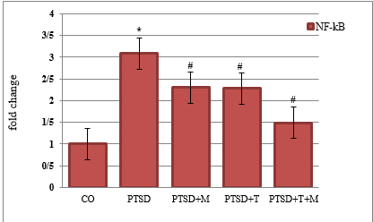
Figure 1. Comparison of NF-κB means in experiences and control groups
*: Significant difference with the control group
#: Significant difference with the PTSD group
Table2. Comparison of the differences between the means of dependent variables in the 5 experimental and control groups
*: p < 0.05
Table3. Significant results of interventions between experimental and control groups in two dependent variables
Table4. Two-way ANOVA results for NF-κB mRNA expression
*: p < 0.05
| Variable | Control | PTSD | Training + PTSD | Milk thistle + PTSD | Concurrent training + milk thistle + PTSD | p |
| Baseline weight (g) | 301 ± 11 | 287 ± 9 | 294 ± 10 | 289 ± 9 | 297 ± 11 | 0.344 |
| Post intervention Weight (g) | 300 ± 9 | 278 ± 8 | 299 ± 9 | 297 ± 7 | 308 ± 11 | 0.027* |
| Weight change (g) | - 1 | - 9 | + 5 | + 8 | + 11 | 0.030* |
| Food intake (g/day) | 23.6 ± 2.1 | 22.8 ± 2.0 | 25.1±2.4* | 24.9 ± 2.3* | 26.3 ± 2.5* | 0.036* |
| Water intake (mL/day) | 31.4 ± 3.2 | 30.5 ± 3.0 | 36.8±3.7* | 35.9 ± 3.5* | 38.1 ± 3.8* | 0.028* |
Figure 1. Comparison of NF-κB means in experiences and control groups
*: Significant difference with the control group
#: Significant difference with the PTSD group
Table2. Comparison of the differences between the means of dependent variables in the 5 experimental and control groups
| Variable | SS | df | MS | F | p | |
| mRNA NF‑κB | Between groups | 5.089 | 4 | 1.272 | 21.48 | 0.0005 * |
| Within groups | 1.421 | 51 | 0.028 | |||
| Total | 6.510 | 55 | ||||
| Serotonin |
Between groups | 5.27 | 4 | 1.317 | 24.51 | 0.0001 |
| Within groups | 2.74 | 51 | 0..54 | |||
| Total | 8.01 | 55 | ||||
Table3. Significant results of interventions between experimental and control groups in two dependent variables
| Groups | NF-κB | Serotonin |
| sig. | sig. | |
| Control vs PTSD | 0.0005* | 0.0001* |
| Control vs Training + PTSD | 0.0001* | 0.0001* |
| Control vs Milk Thistle + PTSD | 0.016* | 0.0002* |
| Control vs concurrent training + milk thistle + PTSD | 0.220 | 0.210 |
| PTSD vs Training + PTSD | 0.520 | 0.180 |
| PTSD vs Milk Thistle + PTSD | 0.580 | 0.001* |
| PTSD vs concurrent training + milk thistle + PTSD | 0.075 | 0.0001* |
| Training + PTSD vs Milk Thistle + PTSD | 0.060 | 0.330 |
| Training + PTSD vs concurrent training + milk thistle + PTSD | 0.002* | 0.0004* |
| Milk Thistle + PTSD vs concurrent training + milk thistle + PTSD | 0.600 | 0.020* |
Table4. Two-way ANOVA results for NF-κB mRNA expression
| Variable | Source of variance | Sum of squares | df | Mean square | Effect size (Partial η²) | Effect size interpretation | F | p |
| mRNA NF-κB | Exercise | 2.600 | 1 | 2.600 | 0.647 | Large | 21.95 | 0.0005* |
| Milk thistle | 2.489 | 1 | 2.489 | 0.636 | Large | 21.01 | 0.0006* | |
| concurrent training + milk thistle + PTSD | 0.000 | 1 | 0.000 | 0.000 | low | 0.00 | 0.9943 | |
| Residual | 1.421 | 51 | - | - | — | - | - |
No significant interaction effect was observed between concurrent exercise training and milk thistle extract (p = 0.994), indicating additive rather than synergistic effects on NF-κB mRNA expression. The changes in serotonin between groups were analyzed by one- way ANOVA (Table 2). One-way ANOVA revealed a significant increase in serotonin in the groups. Tukey's post hoc test showed a significant difference between the means among the 5 experimental and control groups. (Table3)
Serotonin concentrations were significantly reduced in the PTSD group compared to healthy controls, while both combined exercise and milk thistle extract partially restored serotonin levels. The combination of exercise and supplementation (T + M) showed the highest recovery toward control values. (Figure 2)
The results of the two-way analysis of variance (ANOVA) examining the effects of concurrent exercise training, milk thistle extract, and their interaction on serotonin levels are presented in Table 5.
The two-way ANOVA revealed significant main effects of concurrent exercise training and milk thistle extract on serotonin levels (p < 0.05), while their interaction effect was not statistically significant (p = 0.42).
Representative histological images of hippocampal and liver tissues are presented in Figures X–Y. Tissues were stained with hematoxylin and eosin (H&E) to evaluate general morphology, cellular architecture, and potential pathological alterations. Images were captured at 50 μm magnifications to ensure a representative assessment of tissue integrity across experimental groups. Qualitative and quantitative comparisons were made between healthy controls and experimental groups (PTSD, PTSD + milk thistle extract, PTSD + concurrent exercise training, and PTSD + concurrent exercise training + milk thistle extract) to illustrate the effects of the interventions on tissue structure. (Figure 3, 4)
Serotonin concentrations were significantly reduced in the PTSD group compared to healthy controls, while both combined exercise and milk thistle extract partially restored serotonin levels. The combination of exercise and supplementation (T + M) showed the highest recovery toward control values. (Figure 2)
The results of the two-way analysis of variance (ANOVA) examining the effects of concurrent exercise training, milk thistle extract, and their interaction on serotonin levels are presented in Table 5.
The two-way ANOVA revealed significant main effects of concurrent exercise training and milk thistle extract on serotonin levels (p < 0.05), while their interaction effect was not statistically significant (p = 0.42).
Representative histological images of hippocampal and liver tissues are presented in Figures X–Y. Tissues were stained with hematoxylin and eosin (H&E) to evaluate general morphology, cellular architecture, and potential pathological alterations. Images were captured at 50 μm magnifications to ensure a representative assessment of tissue integrity across experimental groups. Qualitative and quantitative comparisons were made between healthy controls and experimental groups (PTSD, PTSD + milk thistle extract, PTSD + concurrent exercise training, and PTSD + concurrent exercise training + milk thistle extract) to illustrate the effects of the interventions on tissue structure. (Figure 3, 4)
Figure 2. Comparison of Serotonin means in experiences and control groups
*: Significant difference with the control group
#: Significant difference with the PTSD group
Table5. Two-way ANOVA results for serotonin levels (ng/g tissue (
| Variable | Source of variance | Sum of squares | df | Mean square | Effect size (Partial η²) | F | p |
| Serotonin level | exercise | 5270.12 | 1 | 3,375.03 | 0.78 | 12.50 | 0.001* |
| Milk thistle | 4800.45 | 1 | 2,400.23 | 0.55 | 8.88 | 0.005* | |
| concurrent training + milk thistle + PTSD | 2300.67 | 1 | 287.58 | 0.30 | 1.06 | 0.420 | |
| Residual | 2740.89 | 51 | 274.09 | — | — | — |
Figure 3. Comparison of H&E-stained hippocampal tissue samples across experimental groups: (A( HC group, (B) PTSD group, (C) PTSD+T group, (D) PTSD+M group, (E) PTSD+M+T group. In healthy controls, hippocampal neurons displayed normal morphology with high cellular density, round or oval nuclei, clear cytoplasmic borders, and a uniform matrix, with minimal abnormalities. In the PTSD group, neurons showed marked pathological changes, including shrinkage, nuclear condensation, apoptosis, and reduced cell density. Hippocampal tissue from PTSD rats subjected to combined exercise showed partial improvement, with scattered neurons, fewer dark nuclei, and signs of increased neurogenesis. Administration of milk thistle extract in PTSD rats resulted in a higher density of round, healthy neurons with larger nuclei and improved structural integrity. The combination of exercise and milk thistle extract produced the most pronounced protective effect, with dense, uniformly organized neurons closely resembling the healthy control group. Hematoxylin and eosin (H&E) staining ×400 magnification. Scale bar = 10 µm.
Figure 4. Comparison of H&E-stained liver tissue samples across experimental groups: (A( HC group, (B) PTSD group, (C) PTSD+T group, (D) PTSD+M group, (E) PTSD+M+T group. In healthy controls, hepatocytes were well-organized with regular, round nuclei, clear cytoplasmic borders, and uniform lobular architecture, showing minimal signs of cellular damage. In the PTSD group, hepatocytes exhibited marked pathological changes, including disorganization, nuclear condensation, cytoplasmic vacuolation, and reduced cell density. Liver tissue from PTSD rats subjected to combined exercise showed partial recovery, with improved hepatocyte arrangement and fewer abnormal nuclei. Administration of milk thistle extract in PTSD rats resulted in higher hepatocyte density, more regular nuclei, and restored lobular structure. The combination of exercise and milk thistle extract produced the most pronounced protective effect, with hepatocytes closely resembling the healthy control group, minimal vacuolation, and uniform tissue architecture. Hematoxylin and eosin (H&E) staining, ×400 magnification. Scale bar = 50 µm.
In healthy control rats, hippocampal neurons and liver hepatocytes displayed normal architecture with intact nuclei, organized cell layers, and no signs of degeneration. In the PTSD group, marked pathological alterations were observed, including neuronal shrinkage, nuclear pyknosis, and disorganized liver hepatocytes, reflecting stress-induced tissue damage.
Administration of milk thistle extract (PTSD + M) partially improved tissue morphology, showing reduced cellular degeneration and more organized hepatocyte and neuronal structures compared with the PTSD group. Concurrent exercise training (PTSD + T) also alleviated histological damage, with hippocampal
neurons and hepatocytes exhibiting improved structural integrity. The combined intervention (PTSD + T + M) demonstrated the most pronounced protective effect, with tissue architecture closely resembling that of healthy controls, corroborating the biochemical findings of elevated serotonin levels and normalized NF-κB expression. These images collectively illustrate the progressive recovery of brain and liver tissue morphology across experimental groups, highlighting the synergistic effects of concurrent exercise training and milk thistle supplementation in mitigating PTSD-induced histopathological alterations.
Discussion
NF-κB mRNA expression was significantly elevated in the PTSD group compared to healthy controls, indicating enhanced activation of this transcription factor in response to traumatic stress. This upregulation was attenuated by both concurrent exercise training and milk thistle extract, with the most pronounced normalization observed in the group receiving both interventions. NF-κB is a key regulator of inflammatory signaling, controlling the transcription of numerous pro-inflammatory cytokines in both central and peripheral tissues. Under stress conditions, NF-κB translocate to the nucleus following IκB degradation, promoting gene transcription associated with inflammation and neuronal stress. Kolb et al., also reported these results (22). Chronic activation of NF-κB in PTSD may contribute to synaptic dysregulation, neuroinflammation, and cognitive impairments. Concurrent exercise training reduced NF-κB expression through anti-inflammatory and antioxidant mechanisms, while milk thistle extract inhibits NF-κB signaling and decreases pro-inflammatory cytokine production (23). Lee et al., reported increased NF-κB activity in immune cells of PTSD patients, and a preclinical study indicates that silibinin, the active component of milk thistle, reduces NF-κB activation and improves behavioral outcomes (24). Some evidence, however, suggests that NF-κB also participates in adaptive neural processes such as synaptic plasticity and memory formation, indicating that its role may be context-dependent (25-26). The results highlight NF-κB as a key mediator of neuroinflammatory responses in PTSD and suggest that concurrent exercise training and milk thistle extract may ameliorate stress-induced pathology through synergistic anti-inflammatory and antioxidant effects (27).
Hippocampal and liver serotonin levels were significantly reduced in the PTSD group compared to healthy controls. Concurrent exercise training and milk thistle extract partially restored serotonin levels, with the combination of both interventions producing the highest recovery toward control values (28). Serotonin, as a key monoamine neurotransmitter, plays an essential role in mood regulation, stress response, and cognitive processes. Chronic stress and traumatic exposure, such as modeled in PTSD, can disrupt serotonin synthesis, reuptake, and receptor signaling, leading to decreased tissue concentrations. The observed decrease in serotonin in PTSD animals is consistent with impaired serotonergic neurotransmission and neuroinflammation. Tseilikman et al., have also reported these findings (29). Exercise enhanced serotonergic signaling, increased tryptophan availability, upregulated serotonin synthesis enzymes, and promoted neurogenesis, particularly in the hippocampus. Milk thistle extract contains bioactive flavonolignans that possess antioxidant and anti-inflammatory properties, which may indirectly preserve serotonergic neurons and enhance serotonin production. Pannu et al., in their review and analysis of pre-clinical studies, the structure activity relationship and characteristics of flavonoids have been found to be useful for clinical trials and the development of antidepressants in terms of therapy (30).
The combined intervention likely exerts synergistic effects. Illesca-Matus et al., and Phillips showed that physical exercise elevates central and peripheral serotonin levels and mitigates stress-induced behavioral deficits (31-32).
Morikawa et al., have reported that serotonergic neurons improved serotonin concentrations in rodent models of stress (33). Contrarily, some studies suggest that the impact of exercise or antioxidants on serotonin may vary depending on stress intensity, duration, or specific brain regions examined, highlighting the context-dependent nature of serotonergic modulation (34).
Administration of concurrent exercise training and milk thistle extract partially restored serotonin levels, with the combination of both interventions producing the highest recovery toward control values. Serotonin, as a key monoamine neurotransmitter, plays an essential role in mood regulation, stress response, and cognitive processes. Strilbytska et al., has pointed out the therapeutic benefits of a number of bioactive substances that have been evaluated in various animal models and human experimental studies. They also report that the anti-anxiety, anti-depressant, and anti-dementia activities of bioactive compounds highlight their potential for the treatment of comorbidities associated with PTSD (35). Chronic stress and traumatic exposure, such as modeled in PTSD, can disrupt serotonin synthesis, reuptake, and receptor signaling, leading to decreased tissue concentrations. The observed decrease in serotonin in PTSD animals is consistent with impaired serotonergic neurotransmission and neuroinflammation )36).
Exercise enhances serotonergic signaling, tryptophan availability, upregulates serotonin synthesis enzymes, and promotes neurogenesis, particularly in the hippocampus. Flavonolignans in milk thistle extract may indirectly preserve serotonergic neurons and enhance serotonin production (37).
Similarly, phytochemicals with antioxidant activity, including silymarin from milk thistle, have been reported to protect serotonergic neurons and improve serotonin concentrations in rodent models of stress (38). Contrarily, some studies suggest that the impact of exercise or antioxidants on serotonin may vary depending on stress intensity, duration, or specific brain regions examined (39-40), highlighting the context-dependent nature of serotonergic modulation.
In the hippocampus, reduced serotonin may contribute to neuronal dysfunction, diminished neurogenesis, and heightened vulnerability to stress-induced apoptosis, while in the liver; it may reflect alterations in peripheral serotonin synthesis, metabolism, and enterohepatic signaling (41). Administration of concurrent exercise training partially restored serotonin levels in both tissues, likely by enhancing tryptophan availability, stimulating serotonin-synthesizing enzymes, and promoting neurogenesis in the hippocampus (42). Milk thistle extract also improved serotonin concentrations, probably through its antioxidant and anti-inflammatory effects, protecting serotonergic neurons from oxidative stress and inflammation-mediated damage (43). The combination of exercise and milk thistle extract produced the most pronounced restoration, with hippocampal and hepatic serotonin levels approaching those of healthy controls (44). demonstrating that physical activity increases central and peripheral serotonin and that antioxidant phytochemicals support serotonergic integrity. Some discrepancies in the literature suggest that the magnitude of serotonin restoration may depend on the type, duration, and intensity of interventions, as well as the specific tissue examined (45).
Overall, the results indicate that concurrent exercise training and milk thistle extract synergistically counteract PTSD-induced serotonergic deficits, supporting improved neuronal function in the hippocampus and normalized peripheral serotonin metabolism in the liver.
Conclusion
Concurrent exercise training and milk thistle (silymarin) administration exerted interactive beneficial effects in male rats with PTSD. The intervention was associated with a reduction in hippocampal NF-κB levels, indicating decreased neuroinflammation, along with an increase in serotonin levels, suggesting improved neurochemical regulation. These findings support the potential of combined exercise and silymarin treatment as a modulator of stress-related neurobiological alterations in PTSD.
Study limitations
First, the sample size per group (n = 11) limits generalizability. Second only male rats were used, and potential sex differences in NF‑κB expression, serotonin levels, and response to interventions were not addressed. Third, molecular and biochemical analyses were limited to hippocampal and liver tissues, while other brain regions or peripheral organs could provide additional insights into systemic effects.
Conflict of interest
The authors declare no competing interests.
Acknowledgements
The authors would like to thank the staff of the animal facility for their assistance in animal care and handling. We also acknowledge the support of the laboratory team in conducting biochemical and molecular analyses.
Funding sources
This research did not receive any specific grant from funding agencies in the public, commercial, or not-for-profit sectors.
Authors' contributions
Conceptualization and Methodology, F.N.; F.S; F.SA; Formal Analysis and Research, F.N.; F.S; F.S.; Writing, Preparation of the original draft, F.N. Writing, Review and Editing, F.N.
Declaration of Interest Statement
The authors declare that they have no known competing financial interests or personal relationships that could have appeared to influence the work reported in this paper.
References
Administration of milk thistle extract (PTSD + M) partially improved tissue morphology, showing reduced cellular degeneration and more organized hepatocyte and neuronal structures compared with the PTSD group. Concurrent exercise training (PTSD + T) also alleviated histological damage, with hippocampal
neurons and hepatocytes exhibiting improved structural integrity. The combined intervention (PTSD + T + M) demonstrated the most pronounced protective effect, with tissue architecture closely resembling that of healthy controls, corroborating the biochemical findings of elevated serotonin levels and normalized NF-κB expression. These images collectively illustrate the progressive recovery of brain and liver tissue morphology across experimental groups, highlighting the synergistic effects of concurrent exercise training and milk thistle supplementation in mitigating PTSD-induced histopathological alterations.
Discussion
NF-κB mRNA expression was significantly elevated in the PTSD group compared to healthy controls, indicating enhanced activation of this transcription factor in response to traumatic stress. This upregulation was attenuated by both concurrent exercise training and milk thistle extract, with the most pronounced normalization observed in the group receiving both interventions. NF-κB is a key regulator of inflammatory signaling, controlling the transcription of numerous pro-inflammatory cytokines in both central and peripheral tissues. Under stress conditions, NF-κB translocate to the nucleus following IκB degradation, promoting gene transcription associated with inflammation and neuronal stress. Kolb et al., also reported these results (22). Chronic activation of NF-κB in PTSD may contribute to synaptic dysregulation, neuroinflammation, and cognitive impairments. Concurrent exercise training reduced NF-κB expression through anti-inflammatory and antioxidant mechanisms, while milk thistle extract inhibits NF-κB signaling and decreases pro-inflammatory cytokine production (23). Lee et al., reported increased NF-κB activity in immune cells of PTSD patients, and a preclinical study indicates that silibinin, the active component of milk thistle, reduces NF-κB activation and improves behavioral outcomes (24). Some evidence, however, suggests that NF-κB also participates in adaptive neural processes such as synaptic plasticity and memory formation, indicating that its role may be context-dependent (25-26). The results highlight NF-κB as a key mediator of neuroinflammatory responses in PTSD and suggest that concurrent exercise training and milk thistle extract may ameliorate stress-induced pathology through synergistic anti-inflammatory and antioxidant effects (27).
Hippocampal and liver serotonin levels were significantly reduced in the PTSD group compared to healthy controls. Concurrent exercise training and milk thistle extract partially restored serotonin levels, with the combination of both interventions producing the highest recovery toward control values (28). Serotonin, as a key monoamine neurotransmitter, plays an essential role in mood regulation, stress response, and cognitive processes. Chronic stress and traumatic exposure, such as modeled in PTSD, can disrupt serotonin synthesis, reuptake, and receptor signaling, leading to decreased tissue concentrations. The observed decrease in serotonin in PTSD animals is consistent with impaired serotonergic neurotransmission and neuroinflammation. Tseilikman et al., have also reported these findings (29). Exercise enhanced serotonergic signaling, increased tryptophan availability, upregulated serotonin synthesis enzymes, and promoted neurogenesis, particularly in the hippocampus. Milk thistle extract contains bioactive flavonolignans that possess antioxidant and anti-inflammatory properties, which may indirectly preserve serotonergic neurons and enhance serotonin production. Pannu et al., in their review and analysis of pre-clinical studies, the structure activity relationship and characteristics of flavonoids have been found to be useful for clinical trials and the development of antidepressants in terms of therapy (30).
The combined intervention likely exerts synergistic effects. Illesca-Matus et al., and Phillips showed that physical exercise elevates central and peripheral serotonin levels and mitigates stress-induced behavioral deficits (31-32).
Morikawa et al., have reported that serotonergic neurons improved serotonin concentrations in rodent models of stress (33). Contrarily, some studies suggest that the impact of exercise or antioxidants on serotonin may vary depending on stress intensity, duration, or specific brain regions examined, highlighting the context-dependent nature of serotonergic modulation (34).
Administration of concurrent exercise training and milk thistle extract partially restored serotonin levels, with the combination of both interventions producing the highest recovery toward control values. Serotonin, as a key monoamine neurotransmitter, plays an essential role in mood regulation, stress response, and cognitive processes. Strilbytska et al., has pointed out the therapeutic benefits of a number of bioactive substances that have been evaluated in various animal models and human experimental studies. They also report that the anti-anxiety, anti-depressant, and anti-dementia activities of bioactive compounds highlight their potential for the treatment of comorbidities associated with PTSD (35). Chronic stress and traumatic exposure, such as modeled in PTSD, can disrupt serotonin synthesis, reuptake, and receptor signaling, leading to decreased tissue concentrations. The observed decrease in serotonin in PTSD animals is consistent with impaired serotonergic neurotransmission and neuroinflammation )36).
Exercise enhances serotonergic signaling, tryptophan availability, upregulates serotonin synthesis enzymes, and promotes neurogenesis, particularly in the hippocampus. Flavonolignans in milk thistle extract may indirectly preserve serotonergic neurons and enhance serotonin production (37).
Similarly, phytochemicals with antioxidant activity, including silymarin from milk thistle, have been reported to protect serotonergic neurons and improve serotonin concentrations in rodent models of stress (38). Contrarily, some studies suggest that the impact of exercise or antioxidants on serotonin may vary depending on stress intensity, duration, or specific brain regions examined (39-40), highlighting the context-dependent nature of serotonergic modulation.
In the hippocampus, reduced serotonin may contribute to neuronal dysfunction, diminished neurogenesis, and heightened vulnerability to stress-induced apoptosis, while in the liver; it may reflect alterations in peripheral serotonin synthesis, metabolism, and enterohepatic signaling (41). Administration of concurrent exercise training partially restored serotonin levels in both tissues, likely by enhancing tryptophan availability, stimulating serotonin-synthesizing enzymes, and promoting neurogenesis in the hippocampus (42). Milk thistle extract also improved serotonin concentrations, probably through its antioxidant and anti-inflammatory effects, protecting serotonergic neurons from oxidative stress and inflammation-mediated damage (43). The combination of exercise and milk thistle extract produced the most pronounced restoration, with hippocampal and hepatic serotonin levels approaching those of healthy controls (44). demonstrating that physical activity increases central and peripheral serotonin and that antioxidant phytochemicals support serotonergic integrity. Some discrepancies in the literature suggest that the magnitude of serotonin restoration may depend on the type, duration, and intensity of interventions, as well as the specific tissue examined (45).
Overall, the results indicate that concurrent exercise training and milk thistle extract synergistically counteract PTSD-induced serotonergic deficits, supporting improved neuronal function in the hippocampus and normalized peripheral serotonin metabolism in the liver.
Conclusion
Concurrent exercise training and milk thistle (silymarin) administration exerted interactive beneficial effects in male rats with PTSD. The intervention was associated with a reduction in hippocampal NF-κB levels, indicating decreased neuroinflammation, along with an increase in serotonin levels, suggesting improved neurochemical regulation. These findings support the potential of combined exercise and silymarin treatment as a modulator of stress-related neurobiological alterations in PTSD.
Study limitations
First, the sample size per group (n = 11) limits generalizability. Second only male rats were used, and potential sex differences in NF‑κB expression, serotonin levels, and response to interventions were not addressed. Third, molecular and biochemical analyses were limited to hippocampal and liver tissues, while other brain regions or peripheral organs could provide additional insights into systemic effects.
Conflict of interest
The authors declare no competing interests.
Acknowledgements
The authors would like to thank the staff of the animal facility for their assistance in animal care and handling. We also acknowledge the support of the laboratory team in conducting biochemical and molecular analyses.
Funding sources
This research did not receive any specific grant from funding agencies in the public, commercial, or not-for-profit sectors.
Authors' contributions
Conceptualization and Methodology, F.N.; F.S; F.SA; Formal Analysis and Research, F.N.; F.S; F.S.; Writing, Preparation of the original draft, F.N. Writing, Review and Editing, F.N.
Declaration of Interest Statement
The authors declare that they have no known competing financial interests or personal relationships that could have appeared to influence the work reported in this paper.
References
- Coleman BL, Gutmanis I, Maunder R, McGeer A. Psychological distress is associated with symptoms of post-traumatic stress disorder among healthcare providers during the COVID-19 pandemic: 2021–2023. Frontiers Psychology. 2025; 16: 1-9.
- Aliev G, Beeraka NM, Nikolenko VN, Svistunov AA, Rozhnova T, Kostyuk S, et al. Neurophysiology and psychopathology underlying ptsd and recent insights into the PTSD therapies-a comprehensive review. Journal of Clinical Medicine. 2020; 9(9): 1-19.
- Girotti M, Bulin SE, Carreno FR. Effects of chronic stress on cognitive function – From neurobiology to intervention. Neurobiology of Stress. 2024; 33: 1-23.
- Sun N, Cui Q, Min M, Zhang M, Liu Z, Wu Y. A new perspective on hippocampal synaptic plasticity and post-stroke depression. The European Journal of Neuroscience. 2023; 58(4): 2961-84.
- Pourhamzeh M, Moravej FG, Arabi M, Shahriari E, Mehrabi S, Ward R, et al. The roles of serotonin in neuropsychiatric disorders. Cell and Molecular Neurobiology. 2022; 42(6): 1671-92.
- Tseilikman VE, Tseilikman OB, Karpenko MN, Traktirov DS, Obukhova DA, Shatilov VA, et al. Unraveling the serotonergic mechanism of stress-related anxiety: focus on co-treatment with resveratrol and selective serotonin reuptake inhibitors. Biomedicines. 2024; 12(11): 2455.
- Govindula A, Ranadive N, Nampoothiri M, Rao CM, Arora D, Mudgal J. Emphasizing the crosstalk between inflammatory and neural signaling in post-traumatic stress disorder (PTSD). Journal of Neuroimmune Pharmacology. 2023; 18(3): 248-66.
- Lawrence S, Scofield RH. Post-traumatic stress disorder associated hypothalamic-pituitary-adrenal axis dysregulation and physical illness. Brain, Behavior, & Immunity-Health. 2024; 41: 1-8.
- Yan M, Man S, Sun B, Ma L, Guo L, Huang L, et al. Gut liver brain axis in diseases: the implications for therapeutic interventions. Signal Transduction and Targeted Therapy. 2023; 8(1): 1-26.
- Ye Q, Yuan S, Cai D. Synergistic potential of natural products and exercise: unveiling molecular mechanisms and innovative therapeutic approaches for liver diseases. Frontiers in Nutrition. 2025; 12: 1-22.
- Gupta S, Guleria RS. Involvement of nuclear factor-κb in inflammation and neuronal plasticity associated with post-traumatic stress disorder. Cells. 2022; 11(13): 1-14.
- Mamun AA, Shao C, Geng P, Wang S, Xiao J. Polyphenols targeting nf-κb pathway in neurological disorders: what we know so far? International Journal of Biological Sciences. 2024; 20(4): 1332-55.
- Zhu M, Chen W, Zhang J. Aerobic exercise, an effective intervention for cognitive impairment after ischemic stroke. Frontiers in Aging Neuroscience. 2025; 17: 1-9.
- Vecchio LM, Meng Y, Xhima K, Lipsman N, Hamani C, Aubert I. The neuroprotective effects of exercise: maintaining a healthy brain throughout aging. Brain Plasticity. 2018; 4(1): 17-52.
- Jaffar HM, Al-Asmari F, Khan FA, Rahim MA, Zongo E. Silymarin: Unveiling its pharmacological spectrum and therapeutic potential in liver diseases—A comprehensive narrative review. Food Science & Nutrition. 2024; 12(5): 3097-111.
- Mukherjee AG, Valsala Gopalakrishnan A. The interplay of arsenic, silymarin, and NF-ĸB pathway in male reproductive toxicity: A review. Ecotoxicology and Environmental Safety. 2023; 252: 1-16.
- Fan Z, Chen J, Li L, Wang H, Gong X, Xu H, Wu l, et al. Environmental enrichment modulates HPA axis reprogramming in adult male rats exposed to early adolescent stress. Neuroscience Research. 2021; 172: 63-72.
- Richter-Levin G, Stork O, Schmidt MV. Animal models of PTSD: a challenge to be met. Molecular Psychiatry. 2019; 24(8): 1135-56.
- Lisieski MJ, Eagle AL, Conti AC, Liberzon I, Perrine SA. Single-prolonged stress: a review of two decades of progress in a rodent model of post-traumatic stress disorder. Frontiers in Psychiatry. 2018; 9: 1-22.
- Alway SE, Paez HG, Pitzer CR. The role of mitochondria in mediation of skeletal muscle repair. Muscles. 2023; 2(2): 119-63.
- Mukhtar S, Xiaoxiong Z, Qamer S, Saad M, Mubarik MS, Mahmoud AH, et al. Hepatoprotective activity of silymarin encapsulation against hepatic damage in albino rats. Saudi Journal of Biological Sciences. 2021; 28(1): 717-23.
- Kolb H, Martin S, Kempf K, Kolb H, Martin S, Kempf K. Traditional health practices may promote nrf2 activation similar to exercise. International Journal of Molecular Sciences. 2025; 26(23): 1-22.
- Sivamaruthi BS, Raghani N, Chorawala M, Bhattacharya S, Prajapati BG, Elossaily GM, et al. NF-κB pathway and its inhibitors: a promising frontier in the management of Alzheimer’s disease. Biomedicines. 2023; 11(9): 1-14.
- Lee B, Choi GM, Sur B. Silibinin prevents depression-like behaviors in a single prolonged stress rat model: the possible role of serotonin. BMC Complementary Medicine and Therapies. 2020; 20(1): 1-12.
- Dresselhaus EC, Meffert MK. Cellular specificity of NF-κB function in the nervous system. Frontiers in Immunology. 2019; 10: 1-14.
- Yang D, Su, J, Chen Y, Chen G. The NF-κB pathway: key players in neurocognitive functions and related disorders. European Journal of Pharmacology. 2024; 984: 1-19.
- Hong C, Schüffler A, Kauhl U, Cao J, Wu CF, Opatz T, et al. Identification of NF-κB as determinant of posttraumatic stress disorder and its inhibition by the Chinese herbal remedy free and easy wanderer. Frontiers in Pharmacology. 2017; 8: 1-17.
- Roberti A, Chaffey LE, Greaves DR. NF-κB signaling and inflammation-drug repurposing to treat inflammatory disorders? Biology (Basel). 2022; 11(3): 1-32.
- Tseilikman VE, Tseilikman OB, Karpenko MN, Traktirov DS, Obukhova DA, Shatilov VA, et al. Unraveling the serotonergic mechanism of stress-related anxiety: focus on co-treatment with resveratrol and selective serotonin reuptake inhibitors. Biomedicines. 2024; 12(11): 1-21.
- Pannu A, Sharma PC, Thakur VK, Goyal RK. Emerging role of flavonoids as the treatment of depression. Biomolecules. 2021; 11(12): 1-49.
- Illesca-Matus R, Ardiles NM, Munoz F, Moya PR, Illesca-Matus R, Ardiles NM, et al. Implications of physical exercise on episodic memory and anxiety: the role of the serotonergic system. International Journal of Molecular Sciences. 2023; 24(14): 1-20.
- Phillips C. Physical activity modulates common neuroplasticity substrates in major depressive and bipolar disorder. Neural Plasticity. 2017; 2017: 1-37.
- Morikawa R, Kubota N, Amemiya S, Nishijima T, Kita I. Interaction between intensity and duration of acute exercise on neuronal activity associated with depression-related behavior in rats. The Journal of Physiological Sciences: JPS. 2021; 71(1): 1-11.
- Rillich J, Stevenson PA. Serotonin mediates depression of aggression after acute and chronic social defeat stress in a model insect. Frontiers in Behavioral Neuroscience. 2018; 12: 1-12.
- Strilbytska O, Koliada O, Lushchak V, Lushchak O. The effects of bioactive compounds on PTSD treatment. Current Neuropharmacology. 2025; 23(10): 1156-68.
- Murrough JW, Czermak C, Henry S, Nabulsi N, Gallezot JD, Gueorguieva R, et al. The effect of early trauma exposure on serotonin type 1B receptor expression revealed by reduced selective radioligand binding. Archives of General Psychiatry. 2011; 68(9): 892-900.
- Klempin F, Beis D, Mosienko V, Kempermann G, Bader M, Alenina N. Serotonin is required for exercise-induced adult hippocampal neurogenesis. The Journal of Neuroscience. 2013; 33(19): 8270-5.
- Ranjan S, Gautam A. Pharmaceutical prospects of Silymarin for the treatment of neurological patients: an updated insight. Frontiers in Neuroscience. 2023; 17: 1-13.
- Clemente-Suárez VJ, Martín-Rodríguez A, Curiel-Regueros A, Rubio-Zarapuz A, Tornero-Aguilera JF. Neuro-nutrition and exercise synergy: exploring the bioengineering of cognitive enhancement and mental health optimization. Bioengineering. 2025; 12(2): 1-59.
- Heijnen S, Hommel B, Kibele A, Colzato LS. Neuromodulation of aerobic exercise—a review. Frontiers in Psychology. 2016; 6: 1-6.
- Paraniak-Gieszczyk B, Ogłodek EA. Neurobiological and existential profiles in posttraumatic stress disorder: the role of serotonin, cortisol, noradrenaline, and IL-12 across chronicity and age. International Journal of Molecular Sciences. 2025; 26(19): 1-30.
- Warner A, Iskander L, Allen K, Quatela I, Borrelli, H, Sachs B. The effects of brain serotonin deficiency on the behavioral and neurogenesis-promoting effects of voluntary exercise in tryptophan hydroxylase 2 (R439H) knock-in mice. Neuro Pharmacology. 2024; 258: 110082.
- Khazaei R, Seidavi A, Bouyeh M. A review on the mechanisms of the effect of silymarin in milk thistle (Silybum marianum) on some laboratory animals. Veterinary Medicine and Science Logo. 2022; 8(1): 289-301.
- Alaca N, Özbeyli D, Uslu S, Şahin HH, Yiğittürk G, Kurtel H, et al. Treatment with milk thistle extract (Silybum marianum), ursodeoxycholic acid, or their combination attenuates cholestatic liver injury in rats: Role of the hepatic stem cells. The Turkish Journal of Gastroenterology. 2017; 28(6): 476-84.
- Basso JC, Suzuki WA. The effects of acute exercise on mood, cognition, neurophysiology, and neurochemical pathways: a review. Brain Plasticity. 2017; 2(2): 127-52.
Type of Study: Research |
Subject:
General
Received: 2026/03/25 | Accepted: 2026/06/9 | Published: 2026/06/20
Received: 2026/03/25 | Accepted: 2026/06/9 | Published: 2026/06/20
References
1. Coleman BL, Gutmanis I, Maunder R, McGeer A. Psychological distress is associated with symptoms of post-traumatic stress disorder among healthcare providers during the COVID-19 pandemic: 2021–2023. Frontiers Psychology. 2025; 16: 1-9.
2. Aliev G, Beeraka NM, Nikolenko VN, Svistunov AA, Rozhnova T, Kostyuk S, et al. Neurophysiology and psychopathology underlying ptsd and recent insights into the PTSD therapies-a comprehensive review. Journal of Clinical Medicine. 2020; 9(9): 1-19.
3. Girotti M, Bulin SE, Carreno FR. Effects of chronic stress on cognitive function – From neurobiology to intervention. Neurobiology of Stress. 2024; 33: 1-23.
4. Sun N, Cui Q, Min M, Zhang M, Liu Z, Wu Y. A new perspective on hippocampal synaptic plasticity and post-stroke depression. The European Journal of Neuroscience. 2023; 58(4): 2961-84.
5. Pourhamzeh M, Moravej FG, Arabi M, Shahriari E, Mehrabi S, Ward R, et al. The roles of serotonin in neuropsychiatric disorders. Cell and Molecular Neurobiology. 2022; 42(6): 1671-92.
6. Tseilikman VE, Tseilikman OB, Karpenko MN, Traktirov DS, Obukhova DA, Shatilov VA, et al. Unraveling the serotonergic mechanism of stress-related anxiety: focus on co-treatment with resveratrol and selective serotonin reuptake inhibitors. Biomedicines. 2024; 12(11): 2455.
7. Govindula A, Ranadive N, Nampoothiri M, Rao CM, Arora D, Mudgal J. Emphasizing the crosstalk between inflammatory and neural signaling in post-traumatic stress disorder (PTSD). Journal of Neuroimmune Pharmacology. 2023; 18(3): 248-66.
8. Lawrence S, Scofield RH. Post-traumatic stress disorder associated hypothalamic-pituitary-adrenal axis dysregulation and physical illness. Brain, Behavior, & Immunity-Health. 2024; 41: 1-8.
9. Yan M, Man S, Sun B, Ma L, Guo L, Huang L, et al. Gut liver brain axis in diseases: the implications for therapeutic interventions. Signal Transduction and Targeted Therapy. 2023; 8(1): 1-26.
10. Ye Q, Yuan S, Cai D. Synergistic potential of natural products and exercise: unveiling molecular mechanisms and innovative therapeutic approaches for liver diseases. Frontiers in Nutrition. 2025; 12: 1-22.
11. Gupta S, Guleria RS. Involvement of nuclear factor-κb in inflammation and neuronal plasticity associated with post-traumatic stress disorder. Cells. 2022; 11(13): 1-14.
12. Mamun AA, Shao C, Geng P, Wang S, Xiao J. Polyphenols targeting nf-κb pathway in neurological disorders: what we know so far? International Journal of Biological Sciences. 2024; 20(4): 1332-55.
13. Zhu M, Chen W, Zhang J. Aerobic exercise, an effective intervention for cognitive impairment after ischemic stroke. Frontiers in Aging Neuroscience. 2025; 17: 1-9.
14. Vecchio LM, Meng Y, Xhima K, Lipsman N, Hamani C, Aubert I. The neuroprotective effects of exercise: maintaining a healthy brain throughout aging. Brain Plasticity. 2018; 4(1): 17-52.
15. Jaffar HM, Al-Asmari F, Khan FA, Rahim MA, Zongo E. Silymarin: Unveiling its pharmacological spectrum and therapeutic potential in liver diseases—A comprehensive narrative review. Food Science & Nutrition. 2024; 12(5): 3097-111.
16. Mukherjee AG, Valsala Gopalakrishnan A. The interplay of arsenic, silymarin, and NF-ĸB pathway in male reproductive toxicity: A review. Ecotoxicology and Environmental Safety. 2023; 252: 1-16.
17. Fan Z, Chen J, Li L, Wang H, Gong X, Xu H, Wu l, et al. Environmental enrichment modulates HPA axis reprogramming in adult male rats exposed to early adolescent stress. Neuroscience Research. 2021; 172: 63-72.
18. Richter-Levin G, Stork O, Schmidt MV. Animal models of PTSD: a challenge to be met. Molecular Psychiatry. 2019; 24(8): 1135-56.
19. Lisieski MJ, Eagle AL, Conti AC, Liberzon I, Perrine SA. Single-prolonged stress: a review of two decades of progress in a rodent model of post-traumatic stress disorder. Frontiers in Psychiatry. 2018; 9: 1-22.
20. Alway SE, Paez HG, Pitzer CR. The role of mitochondria in mediation of skeletal muscle repair. Muscles. 2023; 2(2): 119-63.
21. Mukhtar S, Xiaoxiong Z, Qamer S, Saad M, Mubarik MS, Mahmoud AH, et al. Hepatoprotective activity of silymarin encapsulation against hepatic damage in albino rats. Saudi Journal of Biological Sciences. 2021; 28(1): 717-23.
22. Kolb H, Martin S, Kempf K, Kolb H, Martin S, Kempf K. Traditional health practices may promote nrf2 activation similar to exercise. International Journal of Molecular Sciences. 2025; 26(23): 1-22.
23. Sivamaruthi BS, Raghani N, Chorawala M, Bhattacharya S, Prajapati BG, Elossaily GM, et al. NF-κB pathway and its inhibitors: a promising frontier in the management of Alzheimer’s disease. Biomedicines. 2023; 11(9): 1-14.
24. Lee B, Choi GM, Sur B. Silibinin prevents depression-like behaviors in a single prolonged stress rat model: the possible role of serotonin. BMC Complementary Medicine and Therapies. 2020; 20(1): 1-12.
25. Dresselhaus EC, Meffert MK. Cellular specificity of NF-κB function in the nervous system. Frontiers in Immunology. 2019; 10: 1-14.
26. Yang D, Su, J, Chen Y, Chen G. The NF-κB pathway: key players in neurocognitive functions and related disorders. European Journal of Pharmacology. 2024; 984: 1-19.
27. Hong C, Schüffler A, Kauhl U, Cao J, Wu CF, Opatz T, et al. Identification of NF-κB as determinant of posttraumatic stress disorder and its inhibition by the Chinese herbal remedy free and easy wanderer. Frontiers in Pharmacology. 2017; 8: 1-17.
28. Roberti A, Chaffey LE, Greaves DR. NF-κB signaling and inflammation-drug repurposing to treat inflammatory disorders? Biology (Basel). 2022; 11(3): 1-32.
29. Tseilikman VE, Tseilikman OB, Karpenko MN, Traktirov DS, Obukhova DA, Shatilov VA, et al. Unraveling the serotonergic mechanism of stress-related anxiety: focus on co-treatment with resveratrol and selective serotonin reuptake inhibitors. Biomedicines. 2024; 12(11): 1-21.
30. Pannu A, Sharma PC, Thakur VK, Goyal RK. Emerging role of flavonoids as the treatment of depression. Biomolecules. 2021; 11(12): 1-49.
31. Illesca-Matus R, Ardiles NM, Munoz F, Moya PR, Illesca-Matus R, Ardiles NM, et al. Implications of physical exercise on episodic memory and anxiety: the role of the serotonergic system. International Journal of Molecular Sciences. 2023; 24(14): 1-20.
32. Phillips C. Physical activity modulates common neuroplasticity substrates in major depressive and bipolar disorder. Neural Plasticity. 2017; 2017: 1-37.
33. Morikawa R, Kubota N, Amemiya S, Nishijima T, Kita I. Interaction between intensity and duration of acute exercise on neuronal activity associated with depression-related behavior in rats. The Journal of Physiological Sciences: JPS. 2021; 71(1): 1-11.
34. Rillich J, Stevenson PA. Serotonin mediates depression of aggression after acute and chronic social defeat stress in a model insect. Frontiers in Behavioral Neuroscience. 2018; 12: 1-12.
35. Strilbytska O, Koliada O, Lushchak V, Lushchak O. The effects of bioactive compounds on PTSD treatment. Current Neuropharmacology. 2025; 23(10): 1156-68.
36. Murrough JW, Czermak C, Henry S, Nabulsi N, Gallezot JD, Gueorguieva R, et al. The effect of early trauma exposure on serotonin type 1B receptor expression revealed by reduced selective radioligand binding. Archives of General Psychiatry. 2011; 68(9): 892-900.
37. Klempin F, Beis D, Mosienko V, Kempermann G, Bader M, Alenina N. Serotonin is required for exercise-induced adult hippocampal neurogenesis. The Journal of Neuroscience. 2013; 33(19): 8270-5.
38. Ranjan S, Gautam A. Pharmaceutical prospects of Silymarin for the treatment of neurological patients: an updated insight. Frontiers in Neuroscience. 2023; 17: 1-13.
39. Clemente-Suárez VJ, Martín-Rodríguez A, Curiel-Regueros A, Rubio-Zarapuz A, Tornero-Aguilera JF. Neuro-nutrition and exercise synergy: exploring the bioengineering of cognitive enhancement and mental health optimization. Bioengineering. 2025; 12(2): 1-59.
40. Heijnen S, Hommel B, Kibele A, Colzato LS. Neuromodulation of aerobic exercise—a review. Frontiers in Psychology. 2016; 6: 1-6.
41. Paraniak-Gieszczyk B, Ogłodek EA. Neurobiological and existential profiles in posttraumatic stress disorder: the role of serotonin, cortisol, noradrenaline, and IL-12 across chronicity and age. International Journal of Molecular Sciences. 2025; 26(19): 1-30.
42. Warner A, Iskander L, Allen K, Quatela I, Borrelli, H, Sachs B. The effects of brain serotonin deficiency on the behavioral and neurogenesis-promoting effects of voluntary exercise in tryptophan hydroxylase 2 (R439H) knock-in mice. Neuro Pharmacology. 2024; 258: 110082.
43. Khazaei R, Seidavi A, Bouyeh M. A review on the mechanisms of the effect of silymarin in milk thistle (Silybum marianum) on some laboratory animals. Veterinary Medicine and Science Logo. 2022; 8(1): 289-301.
44. Alaca N, Özbeyli D, Uslu S, Şahin HH, Yiğittürk G, Kurtel H, et al. Treatment with milk thistle extract (Silybum marianum), ursodeoxycholic acid, or their combination attenuates cholestatic liver injury in rats: Role of the hepatic stem cells. The Turkish Journal of Gastroenterology. 2017; 28(6): 476-84.
45. Basso JC, Suzuki WA. The effects of acute exercise on mood, cognition, neurophysiology, and neurochemical pathways: a review. Brain Plasticity. 2017; 2(2): 127-52.
Send email to the article author
| Rights and permissions | |
 |
This work is licensed under a Creative Commons Attribution-NonCommercial 4.0 International License. |